- Article
- Occlusion
Occlusion Fundamentals: The Official Guide for Dentists
Spear Team
•
September 24, 2026
Occlusion touches nearly every decision a dentist makes, from a single composite to a full arch rehabilitation, yet it remains one of the most inconsistently taught topics in dentistry. Occlusion refers to how the upper and lower teeth, the temporomandibular joints (TMJs), and the muscles of mastication function together during contact and movement. Understanding occlusion means understanding all four systems, not just where the teeth touch.
Diagnosing occlusion correctly is part of the clinical foundation Spear teaches across its curriculum, one that reaches more than 20 million patients each year through the advanced skills of Spear-trained dentists and teams. That scale comes from consistency: the same diagnostic approach to occlusion, applied case after case, rather than a different judgment call every time.
This guide covers the fundamentals of occlusion: what it is, how it is classified, how to evaluate it, the appliances used to manage it, and its connection to temporomandibular disorder (TMD), vertical dimension, and craniofacial growth. It’s the Function pillar of Facially Generated Treatment Planning, alongside airway, esthetics, structure, biology (AEFSB), and it shows up in nearly every restorative, orthodontic, and prosthetic decision a dentist makes.
Most dentists learned occlusion as a checklist: adjust the high spot, pick a splint, move on. Spear teaches it as a diagnosis: first, determine whether there is any history of airway-disordered breathing; then identify whether the muscles, joints, and teeth align; and then decide what to treat and when. That sequence, diagnose before you touch a tooth, is what separates predictable outcomes from repeat visits.
What is occlusion in dentistry?
Occlusion is the relationship between four systems working together:
- The teeth
- The temporomandibular joints
- The muscles that move the jaw
- The airway
It’s not a single measurement or a static bite record. When Spear Faculty evaluate occlusion, they’re asking whether all four systems agree or whether one is quietly compensating for the others.
What are the basic components of a stable occlusion?
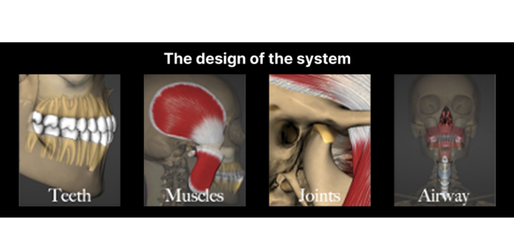
A stable occlusion depends on five things holding true at the same time:
- An airway that supports healthy breathing
- A joint position that the condyles can consistently return to
- Even and simultaneous contact across the posterior teeth in that position
- Anterior teeth that disengage the posterior teeth during excursive movements
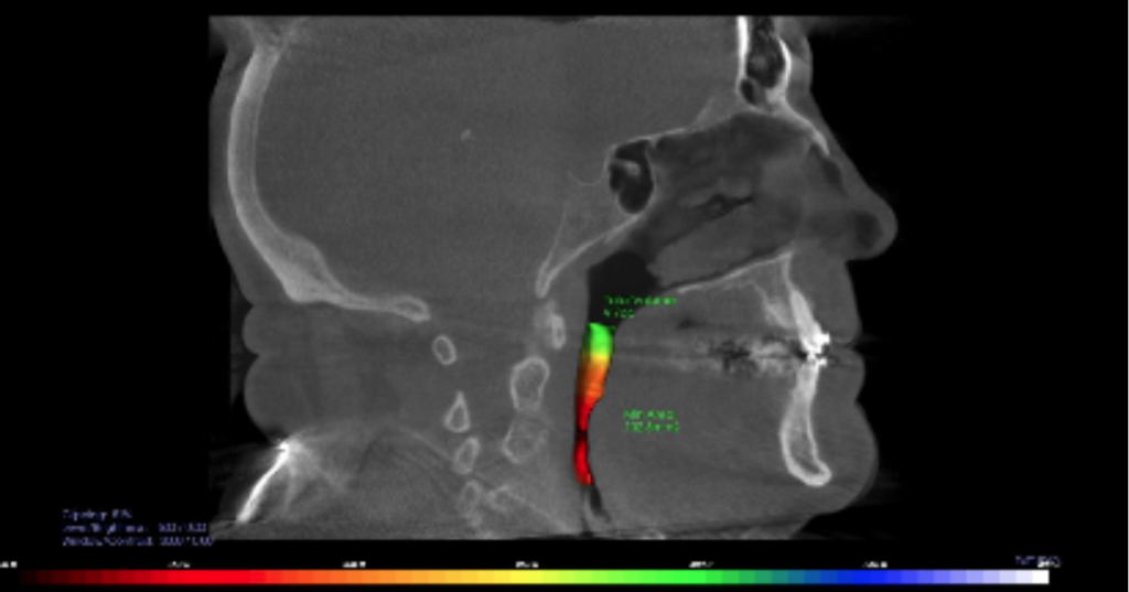
- Muscles that function without excess strain, and an airway that is not driving a compensatory jaw position
When any one of these is missing, the system finds a workaround, often at the expense of a tooth, a restoration, a joint, or nasal breathing.
The occlusal plane is where these five components become visible in a single case. Dr. Gregg Kinzer walks through how to read it and when to change it in The Occlusal Plane Guide. Dr. Carl Steinberg addresses many of the practical questions that come up once dentists start paying attention to these details in Some Questions I’ve Been Asked About Occlusion Over the Years and its follow-up, Common Occlusion Questions, Part 2, including how paper thickness and appliance fit change what a dentist actually sees at the chair.
How is occlusion classified, and what counts as malocclusion?
Occlusion is classified by the anteroposterior relationship of the first molars and canines, first described by Edward Angle in 1899 as Class I, Class II, and Class III. Class I means the molar relationship is normal, regardless of any crowding or rotation. Class II means the lower jaw sits posterior to the upper, which typically presents with increased overjet. Class III means the lower jaw sits anteriorly, presenting as an edge-to-edge or reverse relationship.
Malocclusion is any deviation from that ideal relationship, but the classification alone does not tell a dentist what to treat. A Class I patient can have a destructive occlusion, and a Class II patient can be entirely stable. What matters clinically is whether the classification is forcing a compensation that the restorative plan must live with.
Three patterns come up often enough to warrant their own approach. Excess overjet is covered in Options for Treating Excess Overjet, which outlines when restorative treatment can compensate and when it cannot. Anterior open bite is covered in A Guide to Anterior Open Bites. And a deep overbite in a worn dentition, one of the most common presentations in an adult restorative practice, is covered in How to Fix a Deep Overbite in Wear Patients.
What is centric relation, and why does it matter for occlusion?
Centric relation is the reproducible position of the jaw joints that dentists use as a stable reference point for diagnosis and treatment planning. It matters because a patient’s habitual bite, the position where the teeth happen to come together, is not always the same as their joint-stable position.
That gap first appears in a posterior crown preparation, which Dr. Jeffrey Bonk covers in The Significance of Centric Relation When Preparing Posterior Crowns. As Dr. Jim McKee explains in What Is Centric Relation?, the assumption that the habitual bite and the joint-stable position match holds only when the condyle-disc assembly is functioning normally. Dr. Kinzer walks through the technique for reliably manipulating a patient into centric relation in Demystifying Manipulation for Obtaining Centric Relation, and Dr. Puneet Sandhu’s Centric Relation to Maximum Intercuspation Slide series covers how the gap between centric relation and a patient’s habitual bite develops and why it leads to tooth failure over time.
Centric Relation: a maxillomandibular relationship, independent of tooth contact, in which the condyles articulate in the anterior-superior position against the posterior slopes of the articular eminences; in this position, the mandible is restricted to a purely rotary movement; from this unstrained, physiologic, maxillomandibular relationship, the patient can make vertical, lateral, or protrusive movements; it is a clinically useful, repeatable reference position.
— The Glossary of Prosthodontic Terms 2023, The Journal of Prosthetic Dentistry
How do you evaluate a patient’s occlusion clinically?
A complete occlusal evaluation assesses the airway, the muscles, joints, and teeth in that order before any restorative decision is made. Skipping straight to the teeth is a common reason occlusal problems go undiagnosed until a restoration fails.
Dr. Frank Spear, who developed the diagnostic sequence described in this guide, outlines the muscles every dentist should palpate during a routine exam in Muscle and TMJ Exam: Palpation and Evaluation. Once the exam moves to the joints, Spear’s occlusal exam closely mirrors a recognized diagnostic framework, which Dr. Curt Ringhofer’s breaks down in a two-part series, Diagnosing TMD and Treating TMD: Similarities Between Spear’s Occlusal Exam and the DC/TMD.
Temporomandibular disorders (TMD) are a significant public health problem affecting approximately 5% to 12% of the population. TMD is the second most common musculoskeletal condition (after chronic low back pain), resulting in pain and disability. — Diagnostic Criteria for Temporomandibular Disorders (DC/TMD) for Clinical and Research Applications, Journal of Oral & Facial Pain and Headache
How does occlusion relate to TMD and orofacial pain?
Occlusion and TMD are related but not interchangeable. An occlusal problem can contribute to joint or muscle symptoms, but not every patient with a TMD diagnosis has an occlusal cause, and not every occlusal discrepancy causes pain.
In Why Do Jaws Hurt? Rethinking Orofacial Pain in the 3D Imaging Era, Dr. McKee describes how advances in imaging have moved the profession away from treating jaw pain as a muscle-only problem and toward a more complete picture of joint, disc, and airway involvement. A patient with joint instability needs a different plan than a patient with a purely dental occlusal discrepancy, even when both present with the same symptom. Dr. McKee’s review of which framework to use in Which Joint Classification System Should I Use? covers how to stage what the imaging shows.
How are the TMJ disc and craniofacial growth connected?
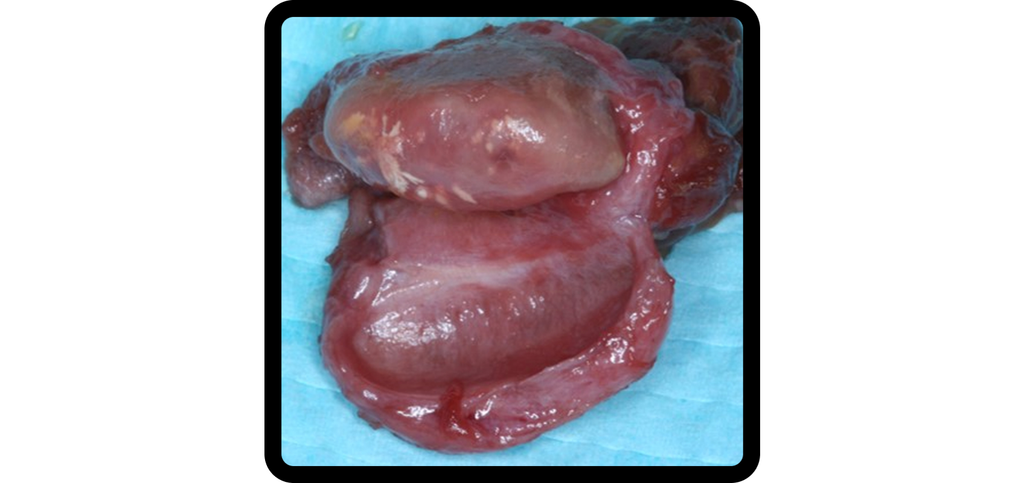
The temporomandibular joint disc plays a direct role in protecting the mandibular condylar cartilage, which functions as the primary growth center of the facial skeleton. When that protective relationship is compromised in a growing patient, skeletal development can shift in ways that extend well beyond clicking or intermittent discomfort.
The Hidden Roles of TMJ Disc Position in Craniofacial Growth explains why disc position, not joint noise alone, should shift a dentist’s diagnostic focus toward developmental risk in younger patients. That connection also shows up in TM Joint Imaging, Part 2: Examining a Clinical Case, where imaging confirms structural changes at the medial pole of a growing condyle, and in Idiopathic Condylar Resorption: Reality or Myth?, which reviews the evidence linking TMJ disc abnormalities to reduced craniofacial growth. Recognizing these signs early changes treatment timing, not just treatment choice.
This study investigated the effects of various treatment modalities for anterior disc displacement on the growth and development of adolescents’ temporomandibular joints. The results suggest that anterior disc displacement treatment can promote condylar growth and reduce the occurrence of maxillofacial deformity. — Impact of treatment modalities on condylar and jaw growth in adolescents with anterior disc displacement of the temporomandibular joint, Journal of Cranio-Maxillo-Facial Surgery
Why does vertical dimension matter in occlusal treatment planning?
Vertical dimension of occlusion is the distance between the upper and lower jaws when the teeth are in contact, and changing it is one of the more consequential decisions in restorative dentistry. Get it wrong, and a patient can end up with muscle strain, speech changes, or esthetic problems that are difficult to reverse.
Dr. Kinzer lays out specific criteria for deciding whether and how much to change a patient’s vertical dimension in Vertical Dimension in Dentistry: Dentate and Edentulous, offering a structured way to evaluate the decision before touching a single tooth.
When is occlusal stability required before restorative treatment?
Occlusal stability should be confirmed before, not after, a dentist commits to a restorative plan. Treating an unstable system can mean having to redo work months or years later, once the underlying instability resurfaces. Dr. Ringhofer addresses this directly in Occlusal Stability in Restorative Dentistry: When Is It Safe To Treat?, outlining TMJ, airway, and craniofacial signs indicating a patient isn’t yet ready for definitive restorative treatment.
A related and common finding, a mandibular slide from centric relation to maximum intercuspation, is covered by Dr. Julie Kwon in Mandibular Slides and Occlusal Management, including how removing a single terminal tooth can destabilize a bite that was already compensating.
What occlusal appliances are available, and how do you choose one?
An occlusal appliance is a reversible way to manage occlusal relationships before committing to permanent treatment, and the options available span a wider range than most dentists use in practice. Occlusal Appliances: The Options walks through the full family of designs, from anterior-only appliances to full-coverage designs built in a seated condylar position, and explains the clinical changes that occur when an appliance is or isn’t built to centric relation.
Choosing among them is a separate question, and the answer is diagnostic rather than habitual. Dr. Ringhofer makes the case for diagnosis-driven selection in The Diagnosis-First Approach to Occlusal Splints. For patients who need anterior repositioning rather than a full-coverage splint, Dr. Jeffrey Rouse describes a less commonly used option in Anterior Bite Guard: The ARA Night Guard You Never Heard Of, and Dr. Kinzer’s The Guide to When and When Not to Splint Restorations lays out the clinical signs that separate the two. For practices moving away from analog impressions, A Digital Approach to Occlusal Appliance Fabrication shows how a fully digital workflow can eliminate chairside adjustment at insertion.
How do bruxism and parafunction affect occlusion?
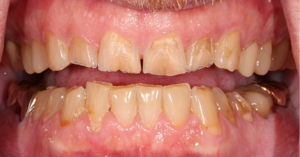
Bruxism and clenching are parafunctional habits, meaning they load the teeth and joints outside of normal chewing function. They do not create an unstable occlusion on their own, but they accelerate what an unstable occlusion would already do. Wear facets, fractured cusps, failing restorations, and muscle soreness are the visible results.
Two things are worth checking before assuming the cause is occlusal. The first is medication. The Link Between Medications and Bruxism covers the drug classes that induce or worsen grinding, which changes the conversation with the patient and their physician. The second is what the wear pattern is telling you. Tooth Wear Appliances: Anterior Bite Planes covers how to protect a worn dentition while the diagnosis is still being confirmed.
What is group function, and is it a problem?
Canine guidance and group function describe two distinct mechanisms by which posterior teeth are protected during lateral movement. In canine guidance, the canine alone disengages the posterior teeth. In group function, several posterior teeth on the working side share the load. Canine guidance is often taught as the ideal, leading dentists to treat group function as something to be corrected. Dr. Kinzer takes that assumption apart in Is Group Function Really Bad? The clinically useful question is not which scheme a patient has, but whether that scheme is causing damage.
What is lingualized occlusion, and when is it used?
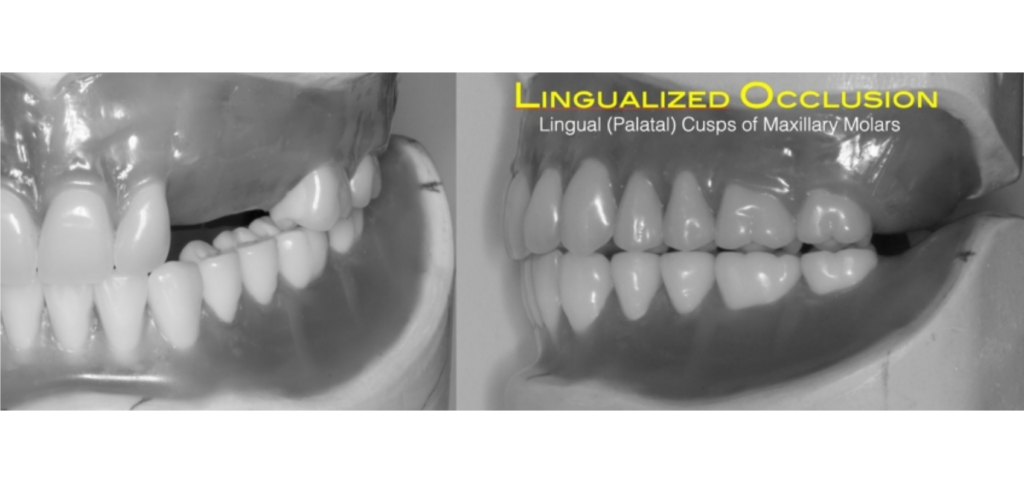
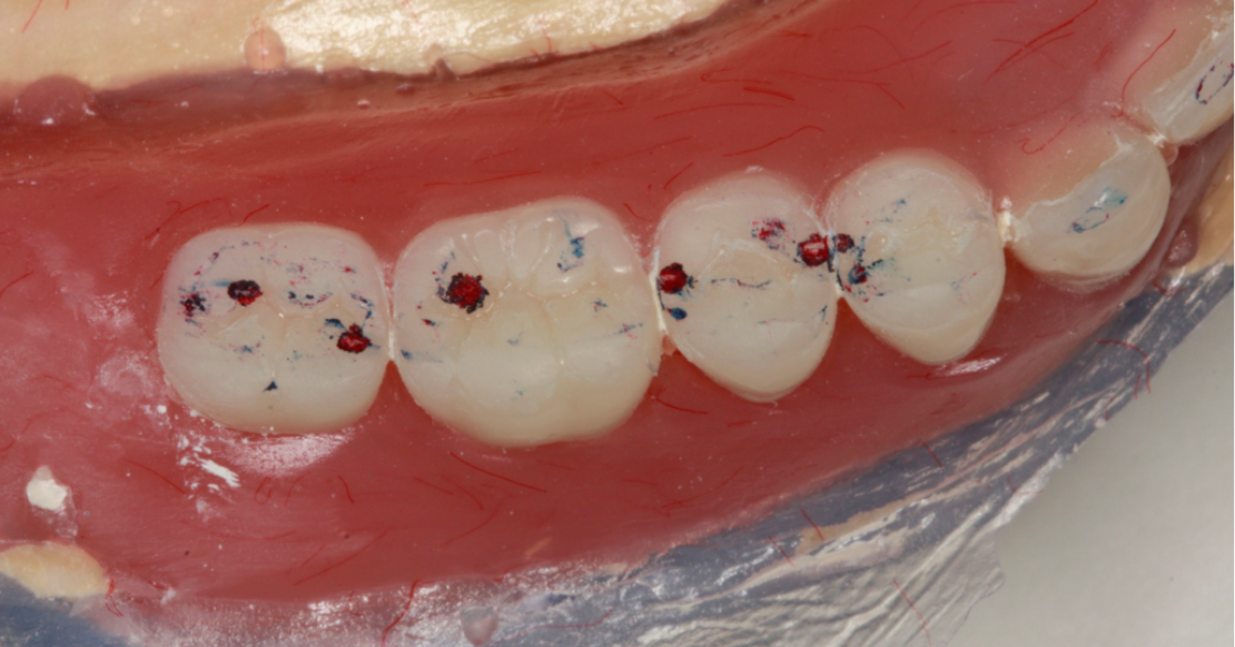
Lingualized occlusion is an occlusal design used for edentulous patients wearing complete dentures. Rather than distributing contact evenly across the posterior teeth, it concentrates contact on the palatal cusps of the maxillary premolars and molars against the central groove of the opposing lower teeth, which reduces lateral interferences and improves denture stability.
Dr. Doug Benting explains the goals and clinical rationale behind this occlusal scheme in Lingualized Occlusion and the Edentulous Patient, including when it makes sense relative to a fully balanced or monoplane design.
Why is a facebow important for restorative accuracy?
A facebow transfers the relationship between a patient’s maxilla and their jaw joints to the articulator, so the lab works from an accurate model of how the patient actually chews rather than a flat guess. Skipping it is a common shortcut after dental school, and an expensive one. Dr. Steve Ratcliff explains what goes wrong without one and how much chairside time a facebow record actually saves in Why a Facebow Is Essential for Restorative Success. For the record itself, 9 Steps to a Facebow Transfer walks through the procedure.
What this means for your practice
A dentist who treats occlusion reactively, adjusting after a fracture rather than diagnosing before one, absorbs a steady cost: avoidable remakes, difficult patient conversations, and cases that take longer than they should. The clinicians who get ahead of this don’t know more facts about occlusion. They diagnose it earlier and more consistently, because occlusion is not an isolated finding. It touches every clinical outcome, from how long a crown lasts to whether a growing patient develops a skeletal discrepancy years later.
If you want to build that diagnostic sequence into how you already practice, Spear’s Occlusion in Clinical Practice Workshop walks through the exact exam and decision-making process covered here, hands-on, with faculty feedback on your own cases. It runs at the Spear Campus in Scottsdale, Arizona, over three days and provides 22.75 hours of ADA CERP and AGD PACE-recognized continuing education credits. If you’d rather start with the online curriculum, Spear Online includes the occlusion pathway on a free trial.
Frequently Asked Questions
SPEAR campus Workshops
Learn Hands-on From Faculty Who Still Practice
A few days at Spear Campus can change how you diagnose, plan, and present cases, not through more lectures, but through hands-on training led by faculty who treat patients every week. Work through real cases in small groups and leave with techniques you can use on your next patient.
Featured Digest articles
Insights and advice from Spear Faculty and industry experts