The Wax Rim Is As Important As Ever!
When considering a component that connects many of the workshops and seminars at Spear Education, it’s the wax rim — or at least the concept of it. This aspect brings everything together, emphasizing precision and detail in dentistry. Let’s explore this fundamental dentistry component, which will remain relevant as new digital workflows emerge.
A fundamental starting point
When Dr. Frank Spear presents his concept of facially generated treatment planning in the workshop Treatment Planning With Confidence, he makes a connection to treating edentulous patients. He refers to how the wax rim is used to identify tooth position, working with facial reference points as a guide. The concept of the wax rim provides an elegant starting point in identifying incisal edge length, midline position, and horizontal plane.
When collecting findings for a patient with apparent signs of worn dentition, the FGTP process includes visualizing a wax rim. Once the teeth have worn, they move to compensate, placing the teeth under consideration for restoration in the wrong place.
A couple of questions arise while figuring out what is possible.
- Would orthodontic tooth movement improve the outcome for the patient?
- Moving ahead with a purely restorative solution decreases treatment time — but at what cost, or what additional treatment may be required?
Moving into the ”prevent, control, resolve” approach championed by Dr. Jeff Rouse in thinking about the airway, the focus at some point in the process becomes the maxillary arch width. The goal is to know how the planned restorative treatment could affect the airway. We don’t want to make it worse, which is where the concept of the wax rim and the connection to FGTP makes another appearance.
Returning to the edentulous patient, the lip support provided by the wax rim has been identified as the starting point before setting maxillary teeth. The L-T-R or “lip, tooth, ridge” concept presented by Drs. Ricardo Mitrani and Darin Dichter begins with lip support, where the goal is to identify facial features for use as a reference point. This approach challenges simply setting the teeth over the edentulous ridge.
Exploring the limitations of a completely digital workflow
One of the challenges in a digital-only protocol for making restorations for edentulous patients arises in the process traditionally reserved for wax rims. The clinical time at this stage aims to facilitate communication with the dental laboratory regarding the desired tooth position. Traditionally, the denture teeth would be positioned on a record base with wax, allowing refinement at the next clinical visit.
A digital workflow has the potential to rely more heavily on a wax rim created with intention. The concept is that the next appointment involves either a printed or milled version for use as either a trial prosthesis or potentially as the definitive prosthesis.

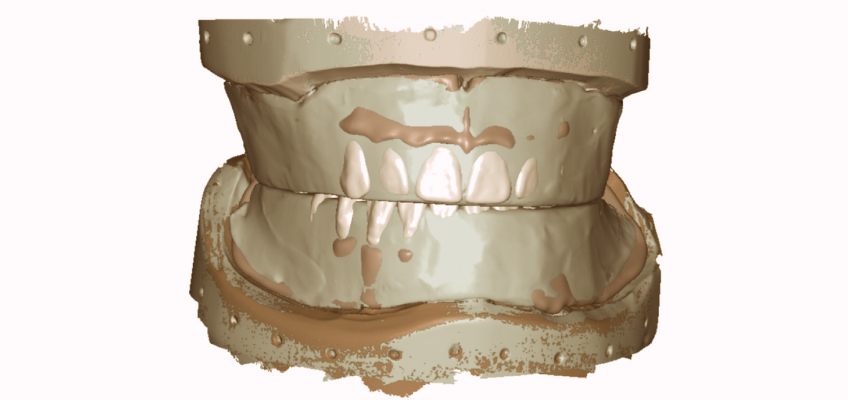
A retentive record base supporting a refined wax occlusal rim is filled with information gained while working with the patient clinically. Once the information is scanned and digitized, it represents an essential connection between analog and digital techniques, because the wax rim represents verified data in three dimensions. Whether the edentulous arches are scanned directly (intraorally) or indirectly (on stone models), selected reference points align the digitized version of the edentulous arch with the digitized version of the wax rim.
- Fang et al.1 describe a technique that combines analog features to create a digital record where the goal is to capture the image of the maxillary edentulous arch, the opposing teeth of the mandibular arch, and the interocclusal vertical dimension of occlusion — all in one clinical visit. A silicone putty is used instead of a wax rim that can be cut back and scanned in segments to capture the information digitally, using the sections as points of reference. A specialized scan retractor is used and placed in the vestibule of the edentulous maxilla, designed with a handle to avoid interference with the scanner.
- Yan and colleagues,2 meanwhile, demonstrate a completely digital workflow working with information derived from CBCT imaging. The patient wore trial dentures with fiduciaries to capture information related to tooth position. The& CBCT as modified to capture traditional cephalometric data points to identify seven landmarks: ANS, PNS, Pog, Me, Go, LGo, and FM. Three angles were measured: PAP (ANS to PNS to Pog), palatal plane (ANS to PNS) to mandibular plane (Me to Go), and facial midline(FM) to AL (ANS to LGo). Working with cephalometric data is interesting; however, a key point here is that the dentures were made previously, and we don’t know if the natural dentition provided the starting point for VDO. How would lip support be verified in this scenario without the existing dentures?
- Ahmed et al.3 describe a complete digital workflow that begins with upper and lower natural dentition back to the second molar. The remaining teeth have to be stable, nonmobile, and in a position close enough to record (allowing for minor modifications) to create an image with enough information to move toward the definitive outcome.
- Papaspyridakos et al.4 present a three-appointment digital workflow with the starting point of existing dentures that are satisfactory to the patient. The interesting point with the all-digital workflow discussion is that the specific step related to the wax rim is noticeably absent. The ideas presented relate more to the concept of the wax rim.
As old-school and analog as it appears, the wax rim — and especially the concept of the wax rim — remains as essential as ever. I’m looking forward to discovering how incorporating the data the wax rim provides advances over time.
References
- Fang Y, Fang JH, Jeong SM, Choi BH. (2019). A technique for digital impression and bite registration for a single edentulous arch. Journal of Prosthodontics, 28(2), e519–e523
- Yan Y, Yue X, Lin X, Geng W. (2023). A completely digital workflow aided by cone beam computed tomography scanning to maintain jaw relationships for implant-supported fixed complete dentures: A clinical study. The Journal of Prosthetic Dentistry, 129(1), 116–124.
- Ahmed WM, Alhazmi A, Alharbi MT, et al. (2023). Maxillary and mandibular complete-arch implant rehabilitation using a complete digital workflow: A case report. Journal of Prosthodontics, 32(8), 662–668.
- Papaspyridakos P, DeSouza A, Bathija A, Kang K, Chochlidakis K. (2021). Complete digital workflow for mandibular full-arch implant rehabilitation in 3 appointments. Journal of Prosthodontics, 30(6), 548–552.
SPEAR ONLINE
Team Training to Empower Every Role
Spear Online encourages team alignment with role-specific CE video lessons and other resources that enable office managers, assistants and everyone in your practice to understand how they contribute to better patient care.

By: Doug Benting
Date: April 9, 2024
Featured Digest articles
Insights and advice from Spear Faculty and industry experts


