Shimstock and Occlusal Maintenance of Dental Implants
Implants are a mainstream and essential treatment choice for the replacement of missing teeth. Maintaining dental implants, like natural teeth, is crucial for promoting health and long-term viability.
All dentists should have a prescribed regimen for evaluating and maintaining dental implants. Patients who get implant treatment should receive systematic and continuous supportive care for the dental implant and the supporting tissues. Ideally, the evaluation of the dental implant should include the following:
- Condition of the soft tissues
- Plaque index
- Tissue probing depth
- Bleeding on probing documentation
- Evidence of suppuration
- Tissue margin position and stability
- Keratinized tissue evaluation
- Mobility and occlusion.
All these aspects should be routinely evaluated at regular re-care or dental checkup appointments. At the checkup, the dentist, hygienist, or another supportive team member can evaluate all criteria and subsequently report any findings to the dentist.
Occlusal Evaluation Is Critical to the Longevity of the Dental Implant
Mucositis is “the reversible inflammation of peri-implant soft tissues.” Alternatively, peri-implantitis is “the irreversible loss of bone support.” Peri-implantitis is a serious condition that requires continuous monitoring and active interception.
As successful as implant therapy has been in clinical practice over the previous 30+ years, there has been a steady increase in peri-implant mucositis and peri-implantitis. Recent studies have determined that the prevalence of peri-implantitis ranges from 1% to 22%.
Current guidelines for maintenance of implant restorations are poorly defined. Most procedures are based upon protocols for patients with natural dentitions. Like I said before, sound guidelines are necessary for evaluating and providing direction toward improving implant outcomes and reducing the incidence of peri-implant disease.
Occlusal evaluation is a critical component for the longevity of the dental implant. Occlusion provides adequate posterior support and eccentric guidance at an appropriate vertical dimension. Potentially destructive effects occur from excursive parafunctional movement and excessive vertical overload. These excessive forces can contribute to rapid and substantial peri-implant bone loss.
The occlusal tactile sensitivity for natural teeth has been measured as low as 8-10 microns. Alternatively, the occlusal sensitivity of dental implants is approximately 48 microns. The lack of periodontal ligament contributes to this delayed response. Bone deformation must occur for an individual to become aware of an occlusal interference. This disparity may easily result in occlusal trauma and a potential for implant periodontitis, bone loss, and subsequent implant loss. Managing occlusal forces is critical for the long-term viability of dental implants.
Here is a suggested protocol for evaluating and managing occlusal forces on dental implants at dental re-care appointments.
It’s common for dentists to check occlusion with articulating papers or ribbon when an occlusal disparity is present. The contact mark left behind allows for visualization of the touchpoint; however, papers and ribbons are too thick relative to the destructive micro forces applied to dental implants. Dental marking ribbons have thicknesses ranging from 20 to 200 microns (depending upon the manufacturer). This significant disparity does not provide adequate information for assessing implant overloading.
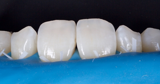
A simple tool and approach to assess implant contact is to use shimstock foil as a tooth/implant contact evaluation tool. Shimstock thickness is on the order of 8-13 microns. It is thinner than articulating papers and ribbons. Although it is not inked and will not leave a mark on the tooth, it provides for a micro-assessment of occlusal holding forces.
Implant patients in my practice are generally placed on a 3- to 4-month re-care cycle. The hygienist implements all the parameters outlined above in evaluating dental implant health and stability. These appointments often alternate between my collaborative implant surgeon/periodontist and our office.
As part of the occlusal evaluation sequence protocol, the dental hygienist places a strip of shimstock foil on the occlusal surface of the dental implant restoration. The patient is instructed to bite together with gentle/regular tooth contact (MIP). While the patient maintains this contact, the dental hygienist attempts to remove the shim stock with cotton forceps.
If the occlusal force is proper, the shimstock should be easily removed with minimal or no resistance. If occlusal contact is present, excess force is applied to the dental implant, and occlusal adjustment is necessary.
The patient should only retain the shimstock when a heavy loading (biting) force is applied. Patients who exhibit holding contact at MIP are scheduled for a subsequent occlusal evaluation and occlusal implant adjustment.
I have found patients are willing to schedule follow-up occlusal adjustment appointments, as they realize the comprehensive evaluation approach and the importance of occlusion and implant longevity.
Patients make significant investments in their dental health. Dental implants are a strategic piece of many dental reconstructions. Maintaining and reinforcing implant longevity is part of our shared objective with patients.
References
- Harper, K. A., & Setchell, D. J. (2002). The use of shimstock to assess occlusal contacts: a laboratory study. International Journal of Prosthodontics, 15(4).
- Sapkota, B., & Gupta, A. (2014). Pattern of occlusal contacts in lateral excursions (canine protection or group function). Kathmandu University Medical Journal, 12(1), 43-47.
- Todescan, S., Lavigne, S., & Kelekis-Cholakis, A. (2012). Guidance for the maintenance care of dental implants: clinical review. Journal of the Canadian Dental Association, 78(1), 107.
- Hogg, K. D. (2017). Implant Maintenance and Review. Practical Procedures in Aesthetic Dentistry, 341-346.
- Weinstein, T., Clauser, T., Del Fabbro, M., Deflorian, M., Parenti, A., Taschieri, S., … & Francetti, L. (2020). Prevalence of peri-implantitis: a multi-centered cross-sectional study on 248 patients. Dentistry Journal, 8(3), 80.
- Derks, J., & Tomasi, C. (2015). Peri‐implant health and disease. A systematic review of current epidemiology. Journal of Clinical Periodontology, 42, S158-S171.
- Forrester, S., Pain, M., Toy, A., & Presswood, R. (2022). THE VALIDITY OF DIFFERENT OCCLUSAL INDICATORS.
- Kim, Y., Oh, T. J., Misch, C. E., & Wang, H. L. (2005). Occlusal considerations in implant therapy: clinical guidelines with biomechanical rationale. Clinical Oral Implants Research, 16(1), 26-35.
SPEAR STUDY CLUB
Join a Club and Unite with
Like-Minded Peers
In virtual meetings or in-person, Study Club encourages collaboration on exclusive, real-world cases supported by curriculum from the industry leader in dental CE. Find the club closest to you today!

By: Jeffrey Bonk
Date: March 10, 2021
Featured Digest articles
Insights and advice from Spear Faculty and industry experts


