Old Tricks for New Dogs: Vertical Margin in Fixed Prosthodontics
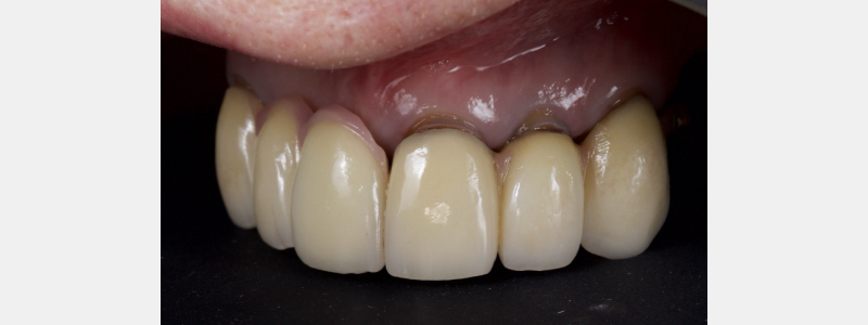
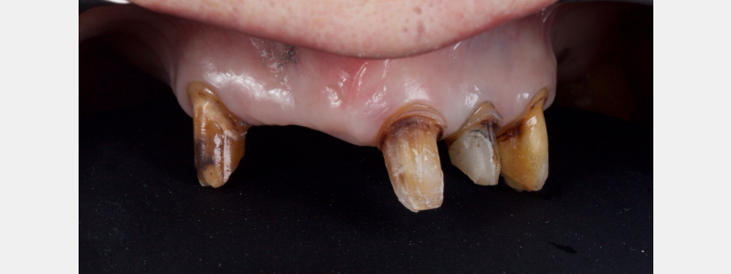
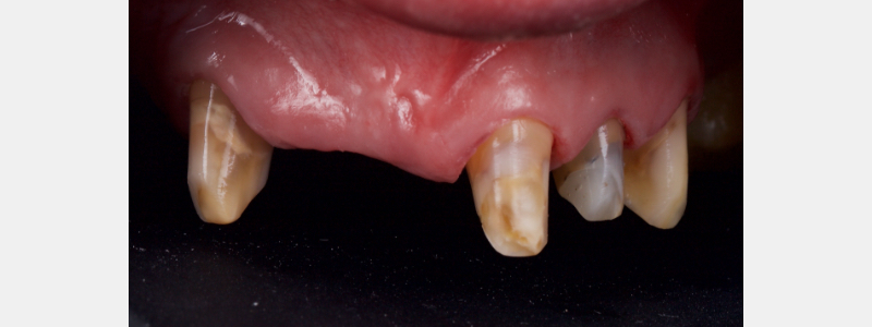
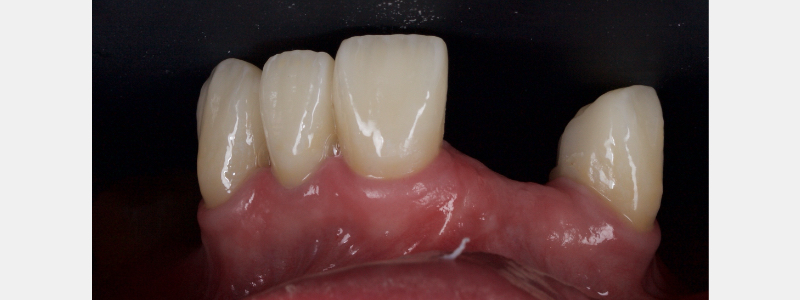
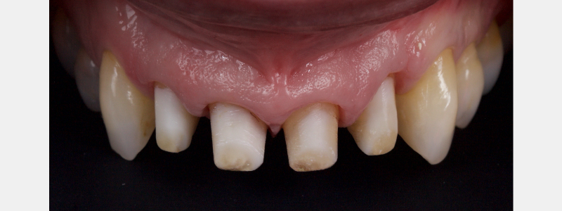
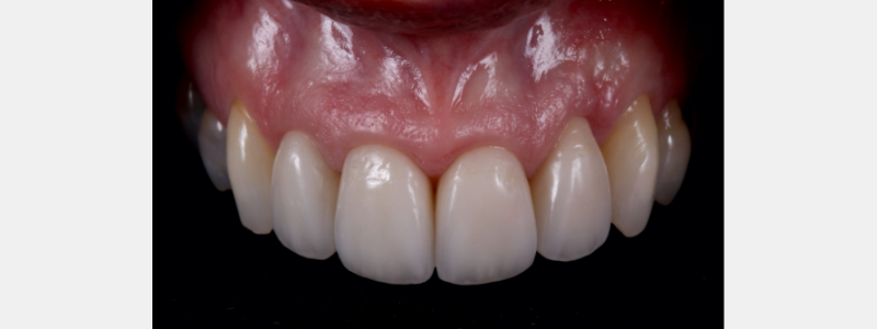
The vertical margin in full veneer crown preparation can offer an esthetic, minimally invasive, and biocompatible solution for periodontally or structurally compromised teeth. For example, this patient, who was restored as a fixed-removable combination case:
The Definition of the Vertical Margin
The finish line is the border between the tooth and the most apical point of the preparation (Castellini, 1994), but the finish line may be:
- Linear Finish Line (sometimes known as “horizontal margin”).
This is where there is a distinct finish line and the clinician defines with a bur the exact position where the restoration should finish on the tooth. The technician then fabricates the restoration to this margin. (Figs. 5 and 6)
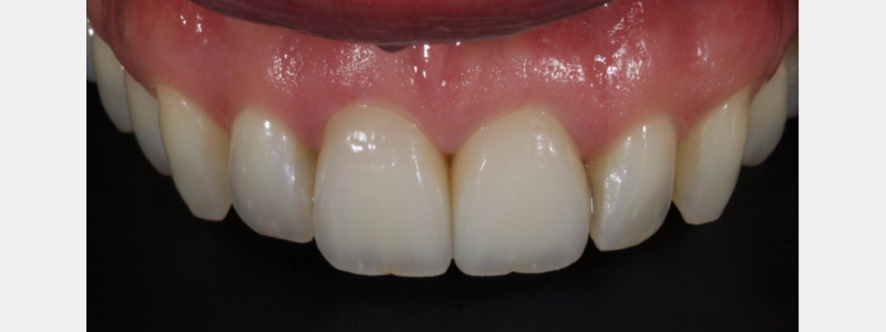
Linear finish lines may be simple (for example, a chamfer or shoulder) or complex (a shoulder with a bevel).
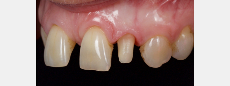
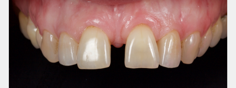
2) Area Finish Line (also known as “vertical margin”).
In this approach, there is a finishing area rather than a line. The clinician defines an area where the crown margin may finish. The clinician and technician then collaborate to determine a finish line position that will give optimal esthetics, biomechanics, and periodontal health. The area finish line may be either feather-edged (where the axial wall meets the root at 180 degrees) or knife-edged (where the axial wall meets the root at less than 180 degrees). (Figs. 7 and 8)
However, a combination of feather and knife edges often coexist on the same prepared tooth, especially if the tooth is multi-rooted.
When Is the Use of the Vertical Margin Advantageous?
Restoration of patients with advanced periodontal disease: When a patient has recession and an exposed root face, finishing at the gingival margin is often desirable to optimize esthetics. However, preparing a root face for a linear margin requires cutting into the root. It may run the risk of devitalization of the tooth or a reduction of the biomechanical performance of the tooth, risking fracture. Therefore, the vertical margin requires no root preparation and is more conservative in these cases.
Restoration of endodontically treated teeth: For the same reason as above the vertical margin does not compromise the horizontal ferrule of root treated teeth.
Provision of splinted crowns: Used to stabilize mobile teeth, multiple units of cross-arch braced crowns can be challenging to prepare with parallel insertion paths. The vertical margin makes this a more straightforward proposition since the technician has some degree of flexibility in margin positioning and can compensate for minor alignment errors.
Preparation of crowns with a previous biologic width invasion: The existing horizontal margin (if minimal depth) can be deleted during the preparation of a vertical margin. Suppose the margin of the provisional crown is placed more coronally than the previous margin. In that case, the tissue can re-establish a healthy biologic width without crown lengthening surgery. This is often advantageous with challenging crown: root ratios.
What Are the Challenges of the Vertical Margin?
Esthetics: this margin design is unsuitable for porcelain-fused to metal crowns since a visible metal margin will result.
Stress-distribution: If not carefully managed, this margin creates high stress distributions compared to other margin types during firing and occlusally loaded (El Ebrashi et al, 1969). This may result in a weak margin in tension and, therefore, be subject to distortion (Kuwata, 1989). Given this, the vertical margin is only suitable for porcelain fused to metal crowns with a metal collar (for non-esthetic zones, e.g., lower molars) or zirconia crowns. Some recent research has also seen success with lithium disilicate.
How Does the Technician Define Margin Position?
Since there is no clearly defined linear finish line, the vertical margin presents a unique challenge: where should the crown margin begin?
If the margin is too coronal, it will be visible and present an esthetic challenge. Conversely, if the margin is placed too apically, it risks impinging on the biologic width, resulting in a periodontal compromise. The solution is straightforward. We will show the approach to this case (Fig. 9).
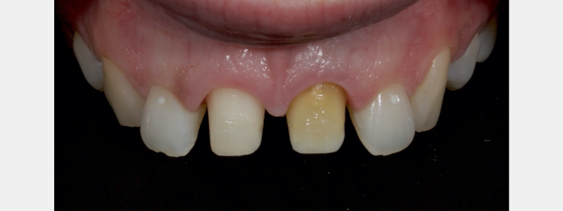
After pouring the dies (or on the scan), prior to trimming the die, the technician marks the position of the gingival margin on the preparation in black pencil (Fig. 10).
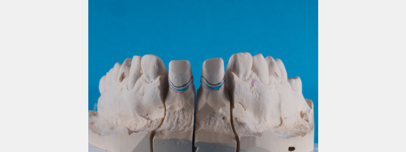
This represents the maximal coronal limit of the potential margin. The die is then trimmed in the conventional manner, and the technician marks the impression’s most apical extension (usually the base of the sulcus) in red pencil.
The finish line can then be created anywhere in the zone between the red and black pencil lines: clearly, in zones where the esthetics are less of an issue (for example, the palatal), the margin is finished closer to the black line.
Where esthetics are more of an issue (for example, discolored substrates)or where creating an ideal emergence contour is more challenging (for example, spaced teeth or peg laterals), you may choose to move the finish line more gingivally.
What Is the Preparation Sequence for Vertical Margins?
The following illustrates an A-Z Stepwise sequence for the preparation of an anterior tooth:
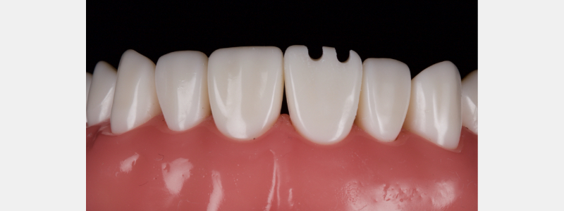
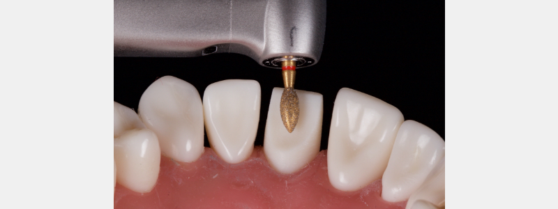
The first phase creates incisal depth grooves for reduction guidance. The workhorse bur for the entire procedure is the 863 016C bur (Fig. 11).
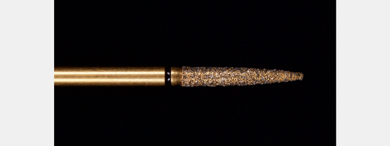
Initially, the portion of the bur closest to the shank, which is a uniform 1.6 mm in diameter, is used at high speed with water spray to create depth grooves at the full diameter of the bur (Figs. 12–14). Caution should be taken not to use the tapered aspect of the bur since this will create non-uniform depth guidance.
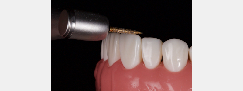
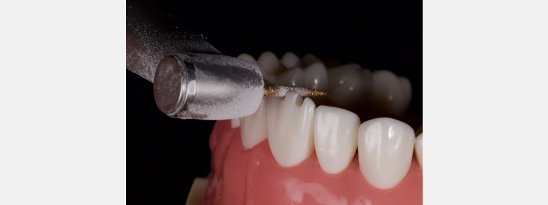
Next, the preparation is separated from the adjacent teeth using an 862 010C burr at medium speed with a water spray (Figure 15): focus should be on the adjacent tooth rather than the tooth being prepared in order to avoid iatrogenic damage.
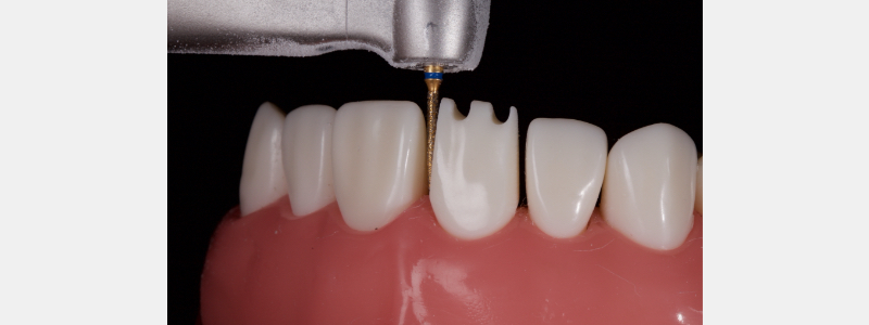
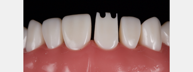
I prefer this bur over a “needle” type design since the flame is much less likely to lose diamond particles and blunt over the needle. The final separation should be slightly over-tapered in terms of Total Occlusal Convergence (TOC) for two reasons: firstly, iatrogenic damage to the adjacent teeth is less likely, and secondly, it should be noted that in the subsequent “gingitage” (Ingraham et al, 1981) stages the TOC tends to reduce as the tooth is prepared. The slight initial over-taper compensates for this and avoids undercuts (Fig. 16).
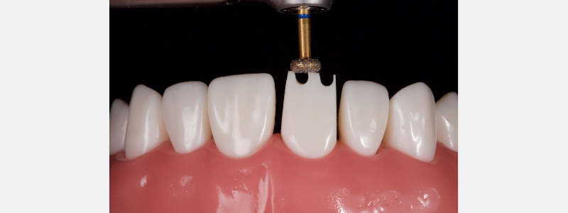
The incisal edge is then reduced to the full depth orientation grooves with a wheel diamond, with my choice being a 909 040 1.5-mm at high speed with water spray (Fig. 17). The wheel is preferred over a cylinder diamond since it offers more control and is less likely to create divots on the incisal edge.
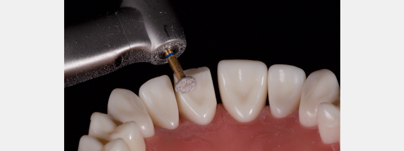
The operator should then revert to the 863 016C bur, using the aspect of the bur closest to the shank to reduce the incisal aspect of the facial surface. Remember, the facial is reduced in two distinct planes to ensure the correct ceramic thickness is available for optimal esthetics (Fig. 19).
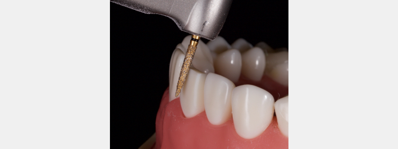
The same bur is then held in the long axis of the tooth to reduce the facial. At this point, the bur should only contact the height of contour, which will be in the mid-third of the tooth (Fig. 20).
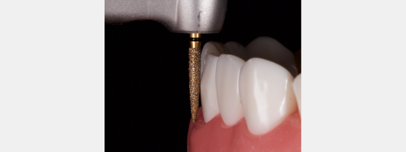
As reduction continues at high speed with water spray, the contact point of the bur gradually moves apically so that finally the bur will be in contact with the tooth at the level of the gingival margin (Fig. 21).
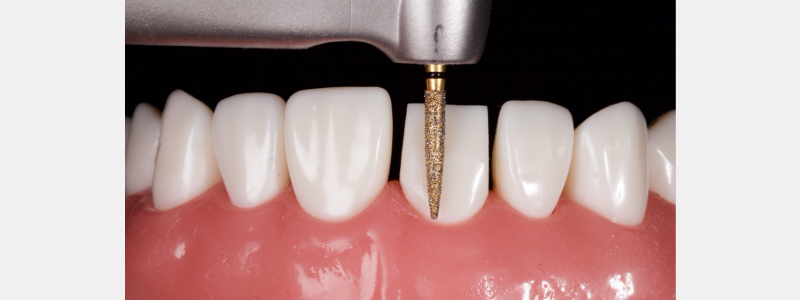
The bur is then worked interproximally and palatally in the same manner until axial reduction is complete. The aim is for 10-20 degrees of taper with a minimum cingulum height of 3.0 mm (Goodacre et al, 2001).
At this point, the progression is toward the “gingitage” phase. The aim is to remove the undercut and create an uninterrupted linear progression from the root surface to the coronal aspect of the prep; further, to remove sulcus epithelium but not connective tissue, to remove chronically inflamed tissue, and to establish a blood clot, which will ultimately recreate an ideal emergence profile.
An 863 012C bur is employed at medium speed with a water spray. The bur is initially advanced at an angle into the sulcus (Fig. 22) and as the undercut is removed, it is slowly inclined into the long axis of the prep (Fig. 23); this ensures the tip of the bur is not cutting and reduces the risk of gouging of the root surface.
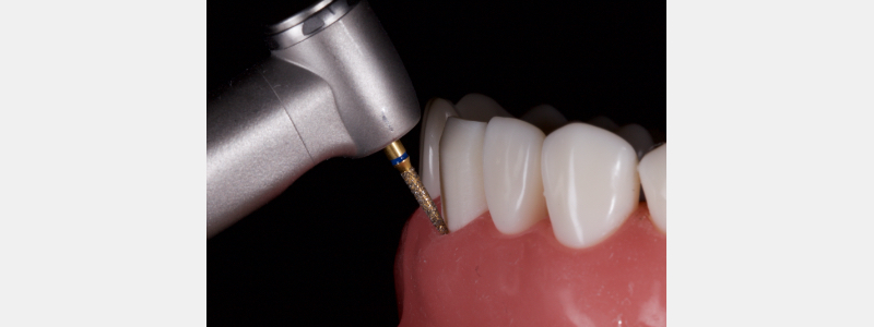
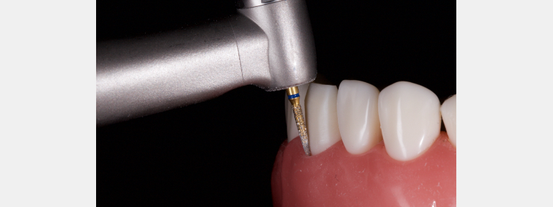
Prior to penetrating the sulcus with the burr it is prudent to assess sulcus depth with a periodontal probe to avoid inadvertent damage to the connective tissue.
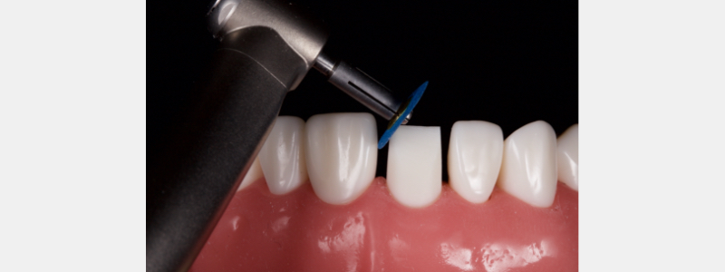
The preparation is then smoothed with fine burrs and discs to create a glassy smooth finish to allow accurate fit of the restoration and soft tissue resolution. Initially, an 863 012F is employed at low speed with water spray within the sulcus (Fig. 24) and then over the reduction planes of the preparation (Fig. 25).
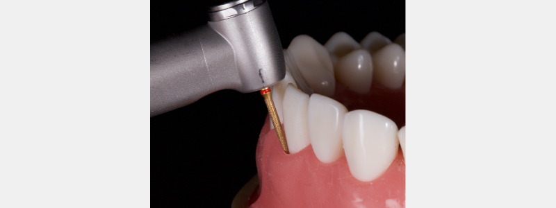
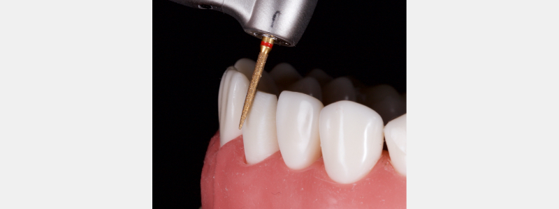
The palatal surface is then smoothed with a 379 023 5.0 mm fine football diamond at very low speed (Fig. 26). Finally, the entire prep is polished with fine discs to create a flowing prep with no sharp line angles (Fig. 27), the outcome (Fig. 28).
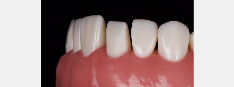
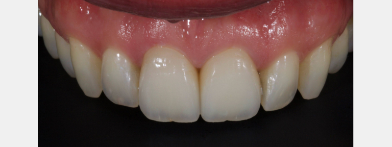
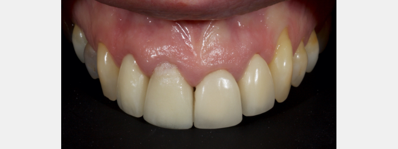
Figures 29–31 show the excellent esthetic and soft tissue outcomes that can be achieved in compromised teeth with vertical margins when zirconia crowns are used in combination.
References
- Castellani, D. (2015). La preparazione dei pilastri per corone in metal ceramica. Bologna: Edizioni Martina.
- El-Ebrashi, M. K., Craig, R. G., & Peyton, F. A. (1969). Experimental stress analysis of dental restorations. Part III. The concept of the geometry of proximal margins. The Journal of Prosthetic Dentistry, 22(3), 333-345.
- Kuwata M. Atlante a colori sulla tecnologia delle riconstruzioni ceramo-metaliche. Utet 1989; 2: 235-250
- Ingraham, R., Sachat, P., & Hansing, F. J. (1981). Rotory Gingival Curettage–A Technique for Tooth Preparation and Management of the Gingival Sulcus for Impression Taking. International Journal of Periodontics & Restorative Dentistry, 1(4).
- Goodacre, C. J., Campagni, W. V., & Aquilino, S. A. (2001). Tooth preparations for complete crowns: an art form based on scientific principles. The Journal of Prosthetic Dentistry, 85(4), 363-376.
VIRTUAL SEMINARS
The Campus CE Experience
– Online, Anywhere
Spear Virtual Seminars give you versatility to refine your clinical skills following the same lessons that you would at the Spear Campus in Scottsdale — but from anywhere, as a safe online alternative to large-attendance campus events. Ask an advisor how your practice can take advantage of this new CE option.

By: Jason Smithson
Date: January 15, 2021
Featured Digest articles
Insights and advice from Spear Faculty and industry experts
