- Article
- Restorative Dentistry
Cracked Cusp Treatment: Essential Guide to 5 Tooth Cracks
Jeffrey Bonk
•
March 5, 2017
Why diagnosing the right crack type is the key to saving a tooth
Editor’s Note: This article has since been revisited and expanded. For the most up-to-date guidance, we recommend reading the latest version by Dr. Jeffrey Bonk.
Cracked cusp treatment starts with a simple question: how much sound tooth structure remains once the fracture is found?
The answer determines whether a tooth can be saved with a direct restoration or needs a crown, an onlay, or even extraction.
What about a cracked tooth? Depending on the crack position and degree, the result may be catastrophic, and a tooth may be lost. This can represent a complete disaster, encompassing emotional, financial, and functional considerations.
The question is: could the cracked tooth have been recognized, and could the outcome have been predicted to avoid a dental catastrophe?
Understanding crack origin, etiology, symptomology, and prognosis supports better diagnosis, patient communication, and timely cracked cusp treatment when a fracture is found.
A cracked tooth is a common occurrence in dentistry; clinicians see them daily in patient treatment. Diagnosing cracks and planning treatment to preserve tooth longevity are critical to helping patients keep their teeth.
One of the primary considerations when a crack is observed is when to intervene. Should the tooth be restored, crowned, or extracted? Identifying and classifying the crack will guide both treatment planning and outcomes. Many cracked teeth can be saved. The keys are identification, understanding signs and symptoms, and early detection.
The American Association of Endodontists has identified five types of tooth cracks: craze lines, fractured cusp, cracked tooth, split tooth, and vertical root fracture. 1, 2
#1: Craze lines: the most common (and least dangerous) tooth crack

Craze lines, also called enamel infractions, are microfractures contained within the enamel only and do not penetrate the dentin layer. All teeth have craze lines, which are more often seen in anterior teeth as vertical striations within the enamel and on marginal ridges. Transillumination provides clear observation of craze lines.
Tooth trauma can contribute to craze lines, whether from blunt force or more recurrent functional forces such as bruxism and parafunction.
Treatment
There are typically no symptoms with craze lines. Treatment can be for esthetic reasons only, and the prognosis is very good. Prevention of bruxism, parafunction, and excessive occlusal trauma is recommended.
#2: Fractured cusp: when a cracked tooth causes biting pain

A fractured cusp is a complete or incomplete fracture of the tooth’s crown that extends subgingivally. The extent and degree of the fractured cusp are variable. The most common cuspal areas to fracture are the lingual cusps of the lower molars and the buccal cusps of the upper molars.
The fracture originates on the occlusal surface and extends gingivally along a buccal or lingual groove and the mesial or distal marginal ridge. Occlusal trauma and force play an integral role in propagating the fracture line. Undermined cusps from existing restorations are also a contributing factor.
The fractured cusp may break and separate at the time of a traumatic event. The resulting tooth segment may be attached to the gingival tissues and may need removal. The remaining exposed tooth area may be sensitive to temperature until restored.
Alternatively, the patient may report biting or temperature sensitivity before the complete cuspal fracture, typically pain on compression and on release of biting pressure. Once the fractured cusp is removed, the biting pain is relieved.
Transillumination can help identify fractured cusps; the light will not penetrate beyond the fractured segment into the rest of the tooth.
Treatment
Depending on the degree of the fracture, there is a good prognosis for retaining the tooth. Root canal therapy or crown lengthening may be needed if the fracture is extensive. Cuspal coverage is recommended for teeth showing early fractured cusp symptoms. Maintaining tooth integrity using crowns or onlays may prevent crack propagation and fracture. Continued, long-term observation is recommended.
#3: Cracked cusp treatment options: planning fractured cusp and cracked tooth restorations
Fractured cusp and cracked tooth cases require individualized cracked cusp treatment planning, since every case carries a different combination of variables.
A fracture can range from a completely asymptomatic finding discovered when an old restoration is removed, to a tooth with constant biting pain and thermal sensitivity, to a fractured cusp with no sensitivity at all. Each presentation calls for a different approach.
Once the extent of the crack and pulp health has been assessed, the first decision point is whether the tooth has a fractured cusp or a cracked tooth.
If it’s a fractured cusp and the pulp is healthy and stable, the fractured segment is typically removed, and the remaining tooth structure is evaluated. If enough sound tooth structure remains, a direct restoration may be sufficient for cracked cusp treatment. If not, the tooth may require an inlay, an onlay, or a build-up and crown.
The choice among these restorative options depends on several factors: the amount of remaining tooth and enamel, the ability to achieve adequate isolation, material selection, whether the restoration will be bonded or cemented, whether the area is esthetically visible, and how the restoration fits into the patient’s broader long-term treatment plan, including adjacent missing teeth, future restorative needs, or anticipated bite or orthodontic changes.
For a true cracked tooth, the crack tends to be more centered in the tooth and, if not managed properly, can progress to a split tooth and eventual tooth loss.
Patients should be informed of the guarded long-term prognosis before treatment begins. If the pulp is irreversibly inflamed or necrotic, root canal treatment is indicated along with an assessment of how deep the crack extends, since deeper cracks carry a greater risk of progressing to a split tooth. If the pulp remains healthy, the goal of treatment shifts to preventing the crack from worsening and sealing it off to prevent bacterial invasion.
The ideal restoration for cracked cusp treatment covers the entire crack while also providing cuspal coverage and support, which is most reliably achieved with a crown or a build-up.
One six-year clinical study3 found that more than 80% of cracked teeth with reversible pulpitis remained vital after being restored with a crown or build-up, with the remaining 20% eventually progressing to irreversible pulpitis.
Some clinicians instead choose more conservative bonded direct or indirect restorations to preserve additional tooth structure; outcome data for these approaches are still developing, but appear promising.4,5 Bonded restorations tend to be more technique-sensitive than crowns and depend heavily on excellent isolation, magnification, and sound bonding principles.
#4: Cracked tooth: diagnosis, symptoms, and treatment challenges

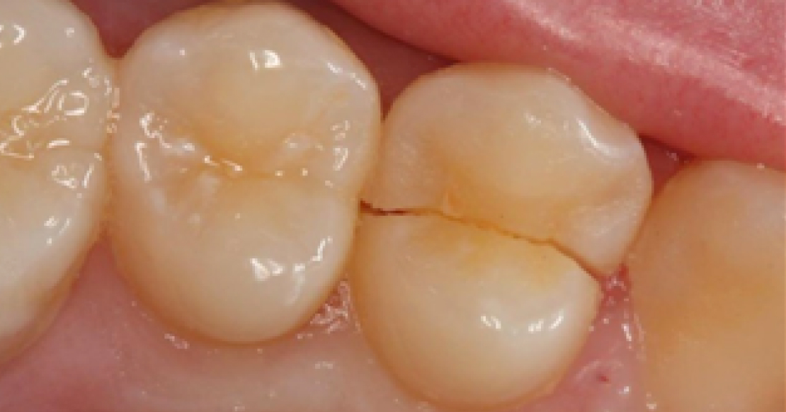
A cracked tooth is defined as an incomplete fracture initiated from the crown and extending subgingivally. The crack is usually oriented mesial to distal. It may extend through one marginal ridge or through both proximal surfaces, and its vertical depth is variable.
The crack may be entirely contained within the crown of the tooth, or it may extend vertically into the root. A cracked tooth is more centered occlusally than a fractured cusp, and because it may progress apically rather than laterally, there is a greater chance of pulpal and periapical pathosis.
The location and extent of a cracked tooth can be difficult to determine. Some cracks are easily seen with magnification or are stained by bacterial migration; others are identified with a dental explorer because they have caused a true separation of the enamel. However, the extent of the surface enamel crack does not directly correlate with the extent of the apical crack. Patient symptoms also vary: some patients experience temperature or biting pain, while others do not.
Excessive occlusal forces are a contributing factor in the development of a cracked tooth. Weakened tooth structure from existing restorations also contributes to tooth cracks. Undermined cusps and marginal ridges create an environment conducive to crack formation. Removal of old restorations is recommended to evaluate the extent and depth of any cracks.
Numerous diagnostic tests are available for cracked tooth situations. Removing old restorations in the presence of a crack is a starting point, and magnification is paramount for assessing the crack’s extent. The crack may be visualized extending along the pulpal floor from mesial to distal; extending the pulpal floor to follow the crack apically can provide information on depth and nerve proximity.
If the crack extends apically into the interproximal area, a perio probe may be used to evaluate for a narrow or isolated band of bone loss vertically down the root, a pathognomonic sign of root fracture. Tooth staining, transillumination, or wedging are additional techniques for assessing the extent of a crack. Pulp vitality and patient symptoms aid in determining the extent of the crack.
Treatment
Cracked tooth treatment is variable and depends on the extent of the crack, the operator’s experience, judgment, and patient symptoms. There are no definitive restorative recommendations in the literature for cracked teeth, so proper diagnosis and preventive strategies are essential.6
Root canal treatment is indicated if pulpal and periapical symptoms dictate, but cracked tooth treatment may be as limited as replacement of a direct restoration or as extensive as full or partial cuspal coverage. The restorative dentist must weigh the extent and depth of the crack against the remaining tooth’s structural integrity to determine the appropriate treatment modality.
Cracked tooth prognosis is always questionable. There is always a possibility that the crack will progress, even with cuspal coverage, since micromovement during tooth function can contribute to long-term crack propagation.
Removing damaging habits, for example, by providing a nightguard and controlling bruxism, covering cusps, and counseling patients on the variability of outcomes, are recommended preventive strategies. Patients should be informed of the questionable prognosis associated with this condition.
H2: #5: Split tooth: when a cracked tooth becomes unsalvageable

A split tooth is a complete fracture initiated in the crown that extends subgingivally, typically through both marginal ridges and proximal surfaces to the proximal root. A split tooth is the result of a cracked tooth, but the segments are now entirely separated. The split may occur suddenly, but it is typically the result of long-term growth from an incomplete crack. Damaging habits such as bruxism, parafunction, and chewing ice can contribute to crack propagation and, ultimately, a split tooth.
Treatment
The split segments may be visualized or identified by wedging them apart, but the prognosis for the tooth is hopeless in most cases. Sometimes only a single root is affected, such as an upper molar root; in those cases, it may be possible to remove the split root and salvage the remaining tooth. Once the tooth is removed, tooth replacement may be discussed and initiated.
Vertical root fracture: the most severe type of cracked tooth

A vertical root fracture is a complete or incomplete fracture of the root in a buccal-to-lingual direction. The fracture may extend the full length of the root or as a shorter segment. There may or may not be associated symptoms, and these fractures are often discovered on routine periapical X-rays.
Virtually all vertical root fractures are associated with a history of root canal treatment. The existence of a sinus tract or a narrow, vertical periodontal pocket along the root surface is consistent with this diagnosis.
Treatment
The prognosis of a vertical root fracture is virtually hopeless in all cases, so prevention is essential. Minimizing dentin removal during root canal therapy improves structural integrity. Avoid posts and post build-ups when possible, reduce condensation forces during obturation, and provide cuspal coverage after root canal treatment.
If you’d like to build even stronger clinical confidence in this area, Spear’s Treatment Planning With Confidence workshop, offered in Scottsdale, dives deep into conservative approaches for stabilizing compromised teeth.
Cracked cusp treatment is a day-to-day decision in dental practices, and getting it right protects both function and long-term tooth survival. Proper diagnosis, paired with the right treatment for each type of crack, gives patients the best chance of keeping their natural teeth for a lifetime.
Contributing Author
Dr. Jeff Lineberry
References
- American Association of Endodontists. Cracking the Cracked Tooth Code. Endodontics: Colleagues for Excellence. 1997 Fall-Winter; 1-13.
- Endodontists, A. A. (2008). Endodontics: Colleagues for Excellence-Cracking the Cracked Tooth Code. Fall-Winter: Chicago, IL, USA.
- Krell, K. V., & Rivera, E. M. (2007). A six-year evaluation of cracked teeth diagnosed with reversible pulpitis: treatment and prognosis. Journal of Endodontics, 33(12), 1405-1407.
- Signore, A., Benedicenti, S., Covani, U., & Ravera, G. (2007). A 4-to 6-year retrospective clinical study of cracked teeth restored with bonded indirect resin composite onlays. International Journal of Prosthodontics, 20(6).
- Opdam, N. J., Roeters, J. J., Loomans, B. A., & Bronkhorst, E. M. (2008). Seven-year clinical evaluation of painful cracked teeth restored with a direct composite restoration. Journal of Endodontics, 34(7), 808-811.
- Lubisich, E. B., Hilton, T. J., & Ferracane, J. (2010). Cracked teeth: a review of the literature. Journal of Esthetic and Restorative Dentistry, 22(3), 158-167.
Frequently Asked Questions
FOUNDATIONS MEMBERSHIP
Build Clinical Confidence In Your First Five Years
The first five years of practice come with decisions that dental school never fully prepared you for. Spear Foundations Membership gives you a full year of practical training, guidance from trusted faculty, and a community of peers working through the same challenges, for less than you’d charge for one crown.
Featured Digest articles
Insights and advice from Spear Faculty and industry experts