- Article
- Treatment Planning
AI in Dental Diagnostics for Stronger Case Acceptance
Mike Skramstad
•
September 1, 2026
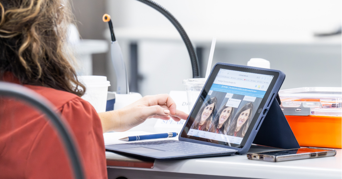
Inside a new patient workflow that puts AI to work from the first visit.
Picture a typical new patient visit. The dentist walks into the hygiene room for the first time, glances at whatever’s been captured so far, and starts piecing together a diagnosis on the spot, while the patient sits there waiting to be told what’s wrong. Everything happens in one narrow window, and there’s no room to miss anything.
Now, picture that same visit with the scans, photos, and radiographs already captured, uploaded, and waiting on the screen before the dentist ever walks in.
That’s what a three-pillar workflow for AI in dental diagnostics makes possible: intraoral scanning, clinical photography, and AI-enhanced radiographs, including CBCT, tied together in the cloud so every exam starts with a complete diagnostic picture already in hand.
Getting there means knowing exactly where AI belongs in that room, and where it doesn’t.
According to the ADA Health Policy Institute’s most recent national survey, 43.3% of dentists already use AI for at least one task in their practice, and another 26.4% plan to do so. Yet, fewer than 5% use it for treatment recommendations, and most have no plans to change that.
Dentists want AI in dental diagnostics to gather and communicate, not to diagnose, and the workflow described below is entirely inside that comfort zone, starting with how the new patient visit itself is restructured to make it possible.
How does AI in dental diagnostics improve the new patient visit?
The foundation of this new workflow is a deliberate restructuring of the new patient appointment.
Every new patient over the age of 16 follows the same protocol. They arrive 30 minutes before their hygiene appointment and spend that time with the dental assistant, who collects all diagnostic information before they see the hygienist or dentist.
This approach accomplishes two things. First, it ensures consistency: when every patient receives the same thorough workup, nothing gets missed. Second, it respects everyone’s time. By the time the dentist walks into the hygiene operatory to meet the patient for the first time, the scans, photographs, and radiographs have already been captured, uploaded, and are waiting on the screen.
Pillar #1: Intraoral scanning with the iTero Lumina
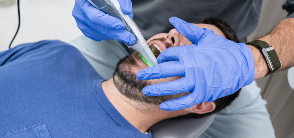
The appointment begins with a full upper- and lower-arch scan using the iTero Lumina intraoral scanner. Many dentists associate the iTero primarily with Invisalign workflows. Still, the Lumina offers something far broader: a diagnostic Oral Health Suite that lets you evaluate tooth health, gum health, occlusion, and overall alignment in three dimensions.
It’s a practical, everyday example of AI-assisted diagnostics: the scanner does the capturing, and the software helps organize what the dentist sees next.
One of Lumina Pro’s standout features is its built-in, high-resolution camera combined with Near-Infrared Imaging (NIRI) technology.
Rather than having the hygienist take a series of individual intraoral photographs, which is time-consuming and often inconsistently executed, the scan itself captures detailed imagery of every tooth surface.
At any point during the patient consultation, the dentist can hover a magnifying tool over any tooth in the scan and pull up a crisp, high-resolution image alongside the NIRI view.
This essentially replaces the need for a separate intraoral photo series during the hygiene exam.
For dental assistants, the Lumina is approachable and efficient. It minimizes the capture of extraneous data, such as tongue and soft tissue, making it straightforward to train nonclinical staff to take accurate, complete scans every time.
That trainability matters beyond convenience. Dental practices nationally are contending with real staffing pressure. About a third of dentists (32.4%) report being overworked, and ongoing shortages, particularly among hygienists, are pushing more non-clinical work onto already-stretched teams.
A workflow that lets assistants reliably capture diagnostic-quality data isn’t just a time-saver. It’s a hedge against a labor market that isn’t loosening up anytime soon.
Pillar #2: A photography protocol for consistency
Photography has historically been the hardest element of a comprehensive new patient exam to implement consistently. The learning curve is real, and most practices that attempt it find themselves taking photos sporadically.
The solution is an assistant-specific photography protocol around a streamlined, abbreviated series. Rather than the full 19-image Facially Generated Treatment Planning series (which remains valuable for advanced case planning), the assistant captures a focused set:
- Portrait
- Retracted portrait
- Full smile
- Lip at rest
- Retracted closed
- Retracted open
- Left and right lateral views
This gives the dentist everything needed to begin a meaningful conversation about esthetics, alignment, and overall dental health.
Camera setup matters. An assistant-specific kit (a compact DSLR body, macro lens, and ring flash) strikes the right balance between image quality and ease of use. For practices just getting started, smartphone-based systems with dental lighting attachments offer an accessible, low-cost entry point.
The key is standardization. When every assistant follows the same script and positions every patient the same way, the photographs become consistent and usable, even if they aren’t the best quality.
Consistent images matter for AI in dental diagnostics, too, since the software driving smile simulations and case presentations works best with clean, predictable source photos.
Pillar #3: Radiographs, CBCT, and where AI changes the conversation
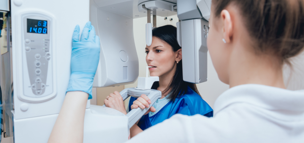
The third and final element of the pre-hygiene data collection is imaging: a full series of intraoral bitewing radiographs plus a CBCT scan. Capturing both before the hygiene visit means the hygienist can focus entirely on the clinical exam and patient relationship, and the dentist enters the room with a complete diagnostic picture already assembled.
CBCT, in particular, has historically been difficult to present to patients. Watching their skull rotate on a screen can be disorienting, and navigating raw CBCT slices in native software doesn’t lend itself to clear patient communication. That’s where AI in dental diagnostics changes the equation, giving the dentist a clearer, three-dimensional starting point for advanced dental treatment planning, rather than a disorienting set of raw slices to interpret on their own.
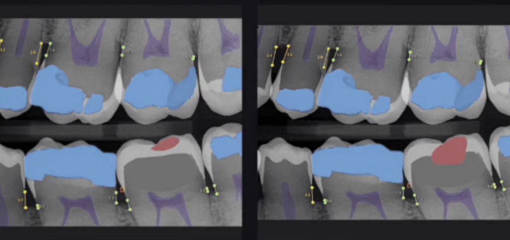
Overjet is an AI-driven radiographic analysis tool that operates on standard bitewing and periapical radiographs. Rather than asking patients to interpret the subtle gray tones of a traditional X-ray, something their eyes aren’t trained to do, Overjet highlights findings in color.
Decay extending into dentin appears in red, areas to monitor in yellow, and existing restorations in blue. A conversation that once required the dentist to describe what they were seeing now becomes visual and immediate.
This distinction mirrors exactly what dentists nationally say they want from AI in dental diagnostics. In the same HPI survey, about one in eight dentists (13.2%) currently use AI to help explain clinical findings to patients. More than two-thirds (68.3%) say they have no plans to use AI that way.
Overjet’s color-coded findings sit on the right side of that gap. The software doesn’t tell the dentist what’s wrong; it gives the dentist a clearer way to show the patient what the dentist has already determined.
As one respondent to HPI’s survey put it, AI “has its place but still must be monitored and verified by humans,” precisely the role Overjet is built for.
Overjet’s CBCT Assist feature extends this capability to three-dimensional imaging. It segments individual teeth and identifies the inferior alveolar nerve and sinuses.
It then presents the information through shortcut views organized by clinical context: implant planning, periodontal evaluation, endodontic assessment, TMJ, airway analysis, and more. The airway module, for example, segments the airway and measures the minimum cross-sectional area, giving dentists a concrete starting point for sleep-related conversations they might otherwise struggle to bring up.
Why is cloud integration essential for AI in dental diagnostics?
The final step of the workflow is where it all comes together: the cloud. All three data streams (scans, photographs, and radiographs) are stored in cloud platforms accessible from any operatory via Google Chrome.
Intraoral scans live in myitero.com. Photographs are uploaded, lightly edited, and organized in DS Core (by Dentsply Sirona). The Canvas feature lets the assistant arrange every image in a consistent layout: one page, all photos visible at a glance, no hunting through folders. Radiographs are analyzed in Overjet’s cloud interface, with both the AI-enhanced bitewings and the segmented CBCT ready to pull up at a moment’s notice.
When the dentist walks in, Google Chrome is already open with three tabs: Overjet, DS Core, and myitero.com. The consultation flows naturally: radiographs first, then photographs, then a deep dive into the 3D scan. Each step builds on the last, giving the patient a clear, visual understanding of their dental health. This is where AI-assisted diagnostics earns its keep: not as a single tool, but as a connected system the whole team can rely on.
What better diagnostics mean for case acceptance
This workflow isn’t about technology for its own sake. It’s about building a system that makes it genuinely easier to show patients what is happening in their mouths and why it matters.
When patients can see decay highlighted in red, visualize their bone levels, or watch a simulation of what orthodontics or restorations could look like, the conversation shifts. Dentistry becomes collaborative rather than directive.
Done consistently, this new patient experience sets the tone for the entire patient relationship. It signals to every new patient, from the moment they arrive, that this practice is thorough, organized, and invested in their long-term care. When the data is complete, the images are compelling, and dental AI is doing the heavy lifting of detection and visualization, treatment acceptance tends to follow naturally.
Practices that lean on AI in dental diagnostics this way aren’t skipping the exam. They’re giving the dentist a head start so that the appointment can focus on the conversation rather than data collection.
How can dentists successfully implement AI in dental diagnostics?
Getting there is as much a training question as a technology question, and it’s rarely a one-person effort. Spear’s course library includes a full series on Overjet itself, plus a broader look at how AI and cloud computing are reshaping diagnosis and case design. A masterclass on AI-assisted removable partial denture design extends these principles further into prosthodontics.
For teams that want to think through where AI in dental diagnostics is headed next, together, rather than one clinician at a time, the Spear Study Club module on AI’s influence in dentistry and the masterclass on the future of AI-powered dentistry are for exactly that conversation. Explore the full lineup through Overjet courses on Spear Online.
The bottom line on AI in dental diagnostics
The technology is here, and the workflow works. AI in dental diagnostics doesn’t replace clinical judgment; it gives dentists better data, clearer images, and more time to spend on the conversation that actually drives treatment acceptance. The question for any practice is simple: have you started?
Frequently Asked Questions
Discover: Overjet Courses
Confidently Step Into AI-Driven Dentistry
The future of diagnostics and treatment planning is already here. Build confidence with AI-powered tools from structured clinical training within Spear Online.
Featured Digest articles
Insights and advice from Spear Faculty and industry experts