Three Solutions for Congenitally Missing Lateral Incisors: A Photo Essay
Addressing congenitally missing lateral incisors is a common condition that justifies an interdisciplinary solution, such as canine substitution, resin-bonded single retainer bridge (resin-bonded fixed dental prostheses or RBFDP), cantilever bridge, or a single-tooth implant. However, delivering the most efficient and predictable solution can be challenging based on a patient’s specific situation, such as age, space availability, hard and soft tissues, finances, and patient preference. This photo essay covers the first three of the four solutions. An upcoming article will discuss the fourth solution for addressing congenitally missing lateral incisors, a single-tooth implant.
Solution #1: Canine Substitution for Missing Lateral Incisors
Canine substitution is the least invasive option and a popular alternative, but it often poses esthetic and functional challenges that need to be considered and cleared (Fig. 1). From an esthetic standpoint, the canine’s shape and shade must be considered. From a contour standpoint, the width of the canine should be evaluated because they are generally larger than lateral incisors.
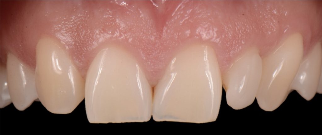
However, the most critical aspect to manage is the CEJ width because it cannot be narrowed. The wider the tooth at the CEJ, the more difficult it is to make a canine look like a lateral incisor. Moreover, canines typically present with a very distinctive root eminence, and if it is particularly accentuated, it could become yet another esthetic challenge — one commensurate with the patient’s lip mobility.
There is no major esthetic concern for patients where a low lip line conceals the gingival outline. Still, if there is high lip mobility and the gingival outline is not concealed, such an eminence could represent an unacceptable esthetic problem.
From a shade standpoint, canines usually are the teeth with the most saturated chroma in the maxillary arch, which often creates an esthetic challenge where this oversaturation is evident.
Consequently, considering these aspects, the ideal clinical scenario for canine substitution would be in patients with smaller shaped canines that are not oversaturated with chroma and in patients who display low lip mobility.
Solution #2: Resin-Bonded Fixed Dental Prostheses (RBFDP)
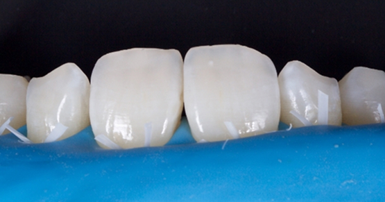
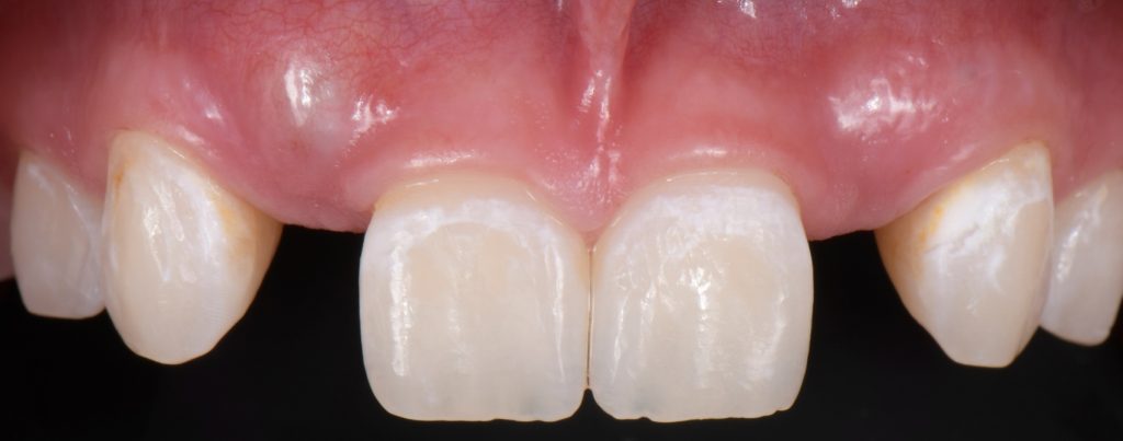
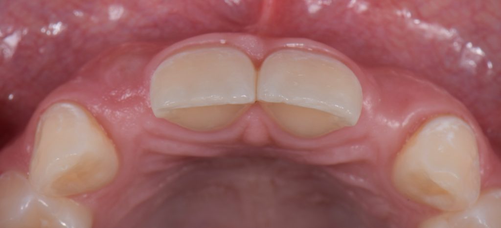
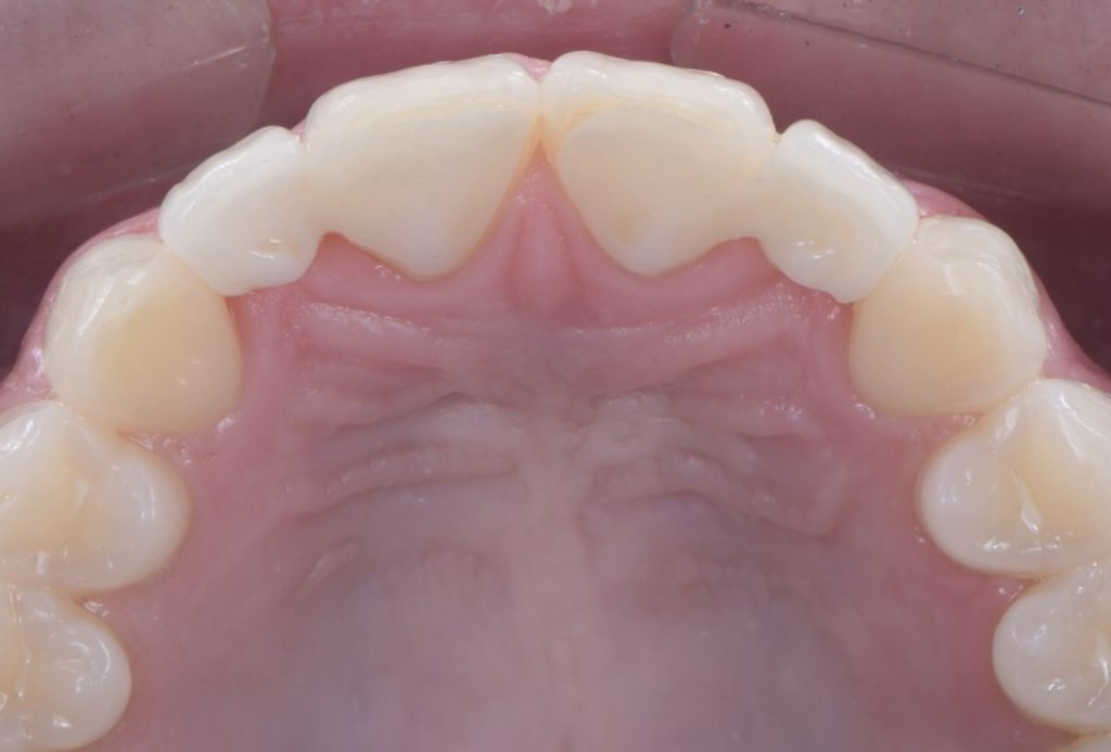
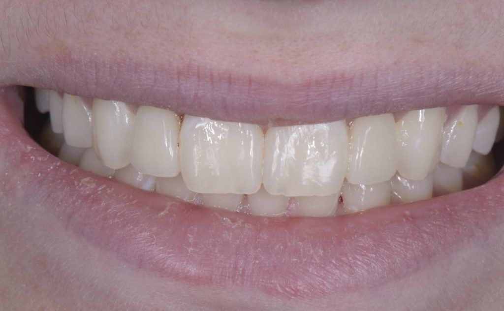
RBFDP is a proven solution for congenitally missing lateral incisors (Figs. 2-12). Although it is considered an interim restoration, the literature provides substantial evidence supporting its long-term potential. However, the clinical performance of an RBFDP is significantly superior to that of a bilateral retainer, and the dissimilar mobility of the abutment teeth explains this.
When placing an RBFDP from a central incisor to a canine, each abutment wants to move under occlusal load, but because of the position each tooth occupies in the arch, loading occurs in different vectors, therefore, leading to debonding of the retainer of the abutment tooth with the least mobility. When considering which one of the adjacent teeth will work best as the abutment, the clinician needs to evaluate:
- Which tooth has more enamel?
- Which tooth has more interocclusal clearance?
- Which tooth is exerting less functional load?
From an occlusal standpoint, patients with shallow overbites or a large amount of overjet make better candidates for RBFDP, and the pontics should be avoided in all lateral excursions, including crossover.
Space requirements and connector dimensions depend on material selection. Utilizing zirconia has been proven to be more predictable over time, and recommended connector dimensions are:
- 3.0 mm in height
- 2.0 mm in width
- The retainer wing thickness is 0.7 mm
The amount of tooth reduction is based on available interocclusal space, and often, there is enough space requiring minimal preparation. The key is to stay in enamel. It is also advisable to stay 2.0 mm away from the incisal edge so that the zirconia retainer does not affect the translucency of the natural tooth.
If the patient has a deep overbite, proclined teeth, or mobile abutment teeth, an RBFPD may not be the best treatment option. This is why they are often used as a long-term provisional until the patient is old enough to have an implant placed.
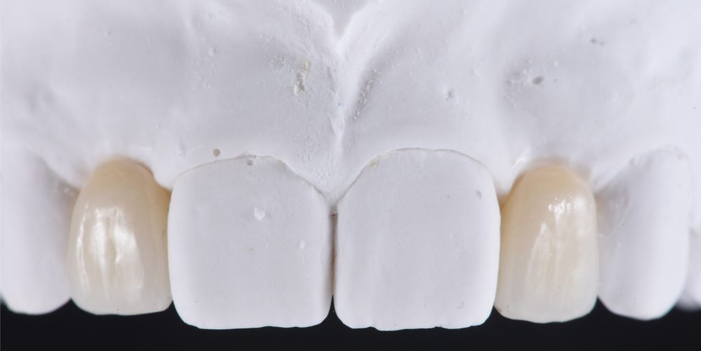
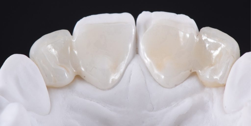
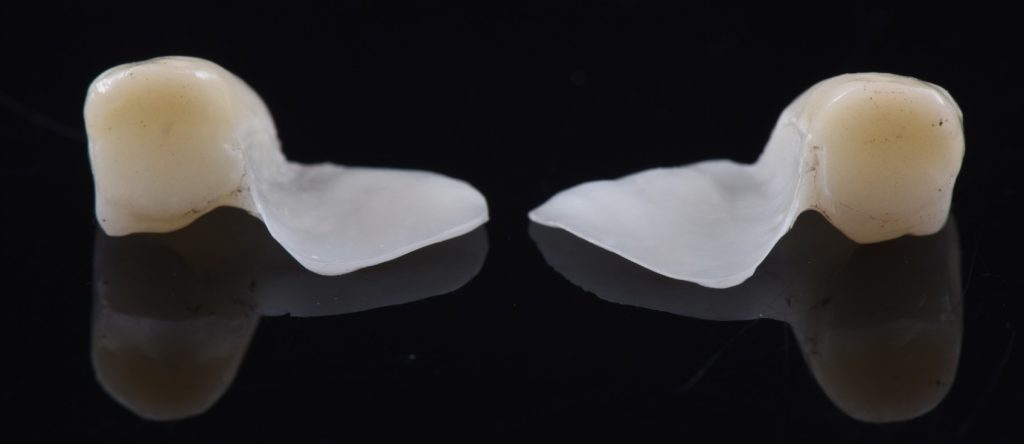
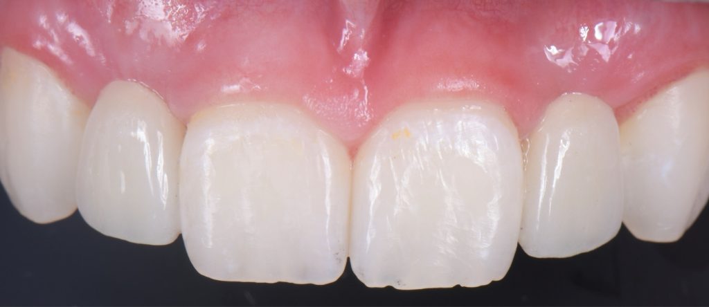
Solution #3: Cantilever Bridge
A fundamental principle in contemporary therapy is to conserve tooth structure by being as minimally invasive as possible. Today, doing a full-coverage restoration on intact natural teeth is extremely difficult to justify, but there are specific conditions where a cantilever bridge can be considered a choice solution.
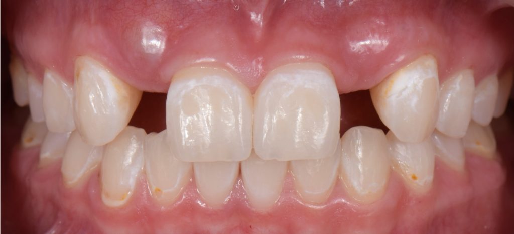
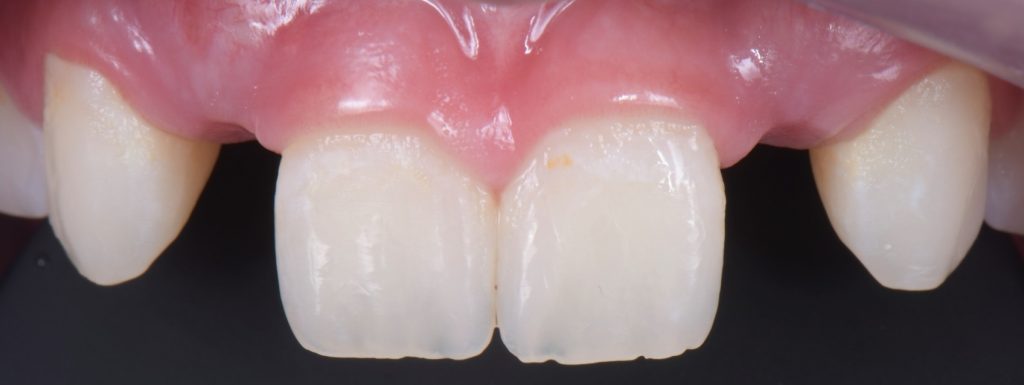
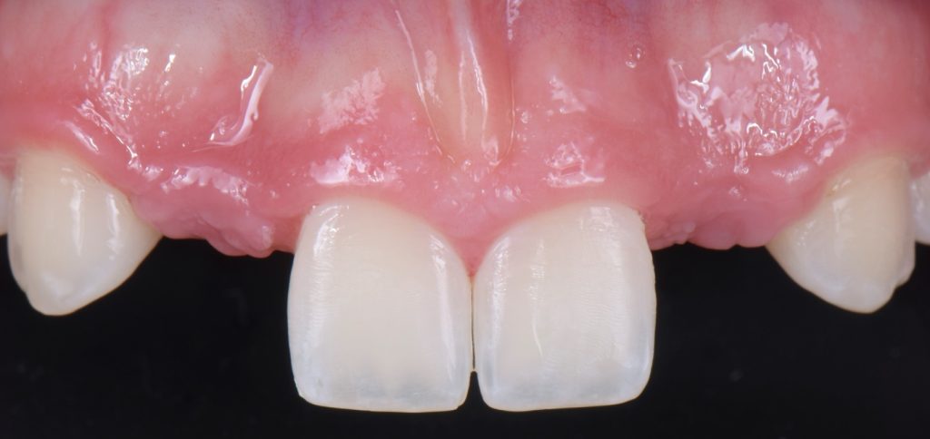
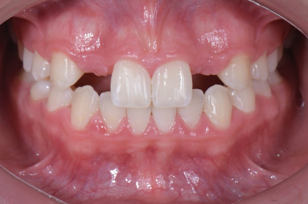
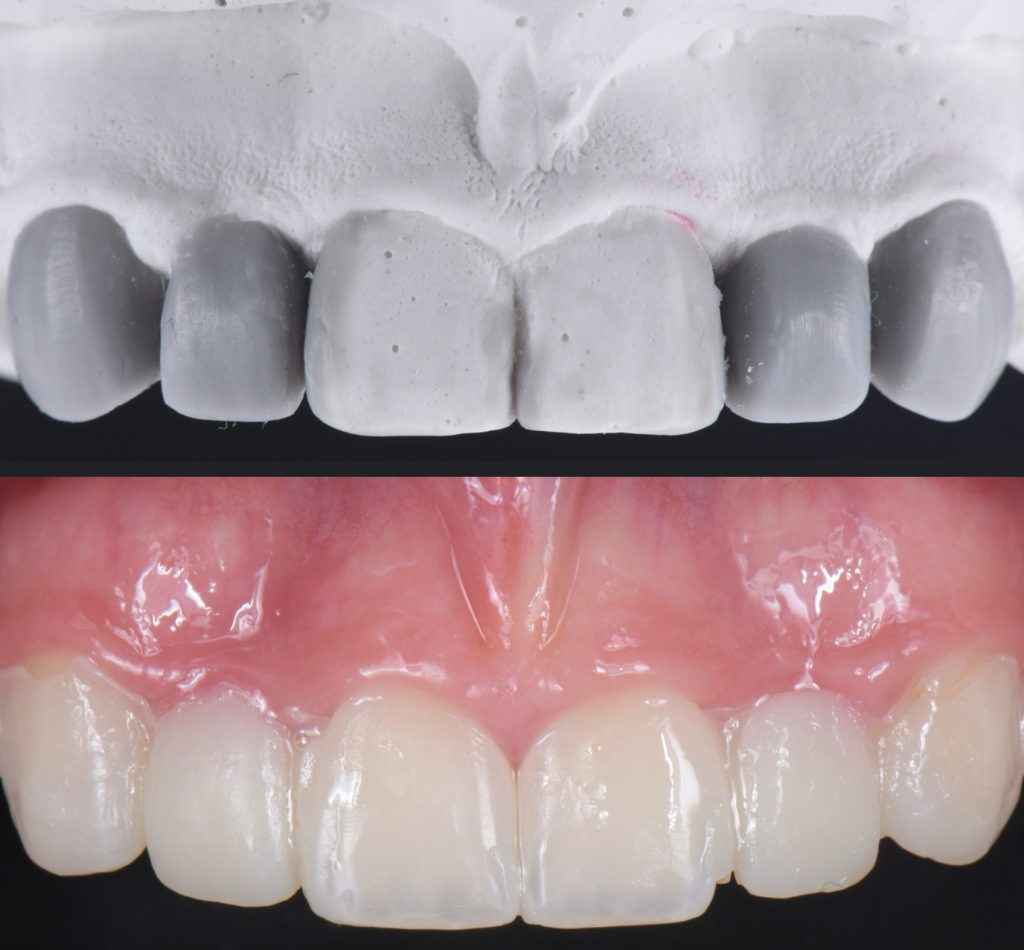
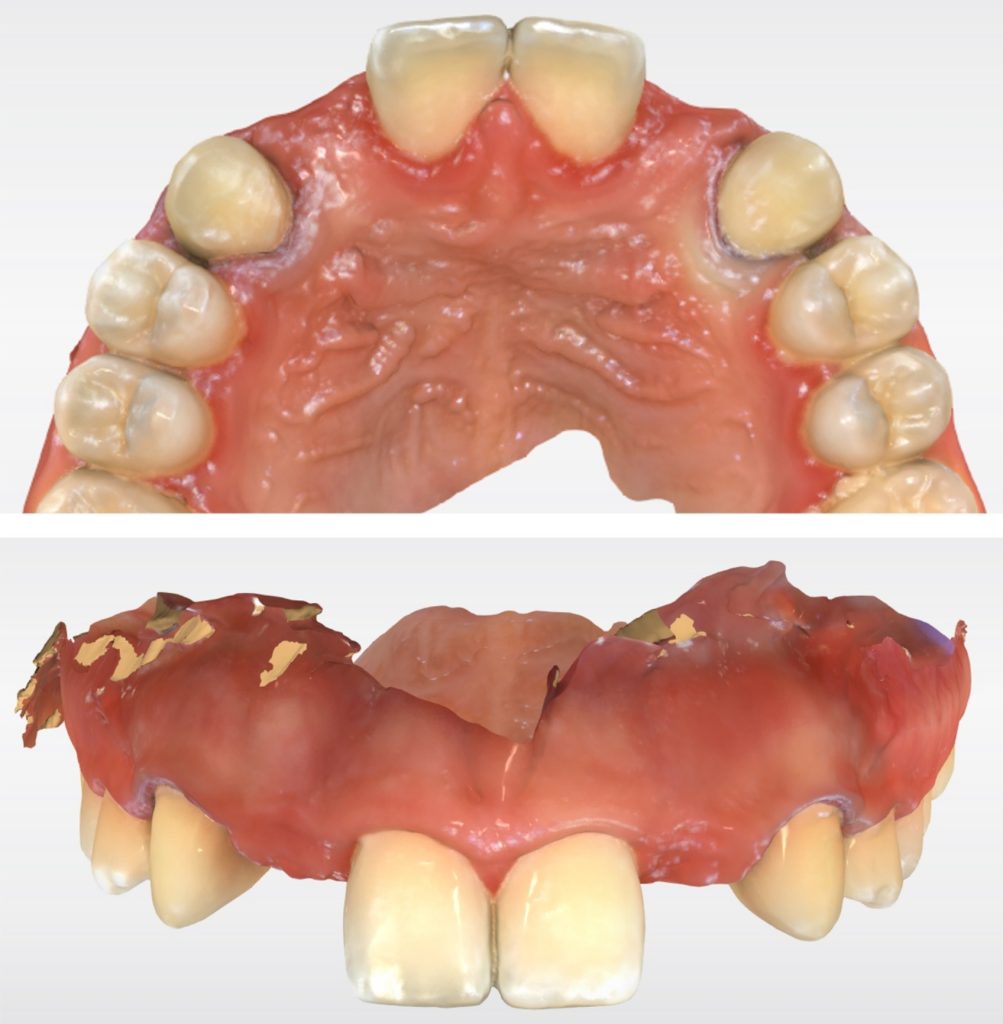
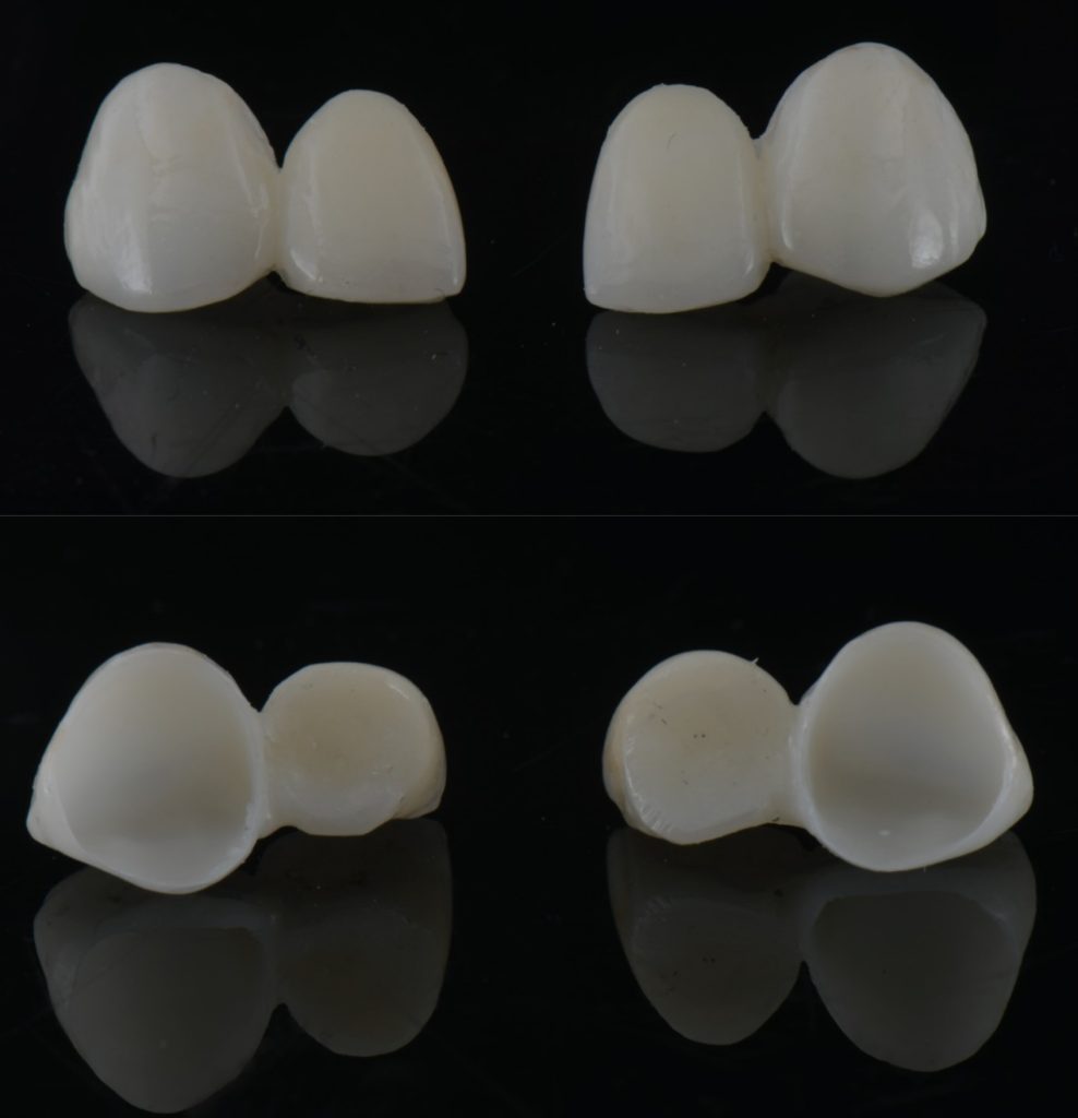
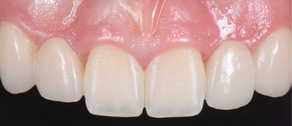
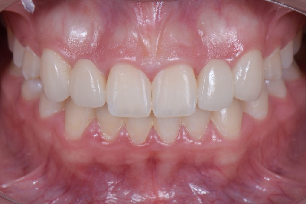
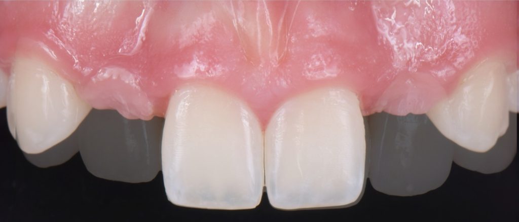
In this case, a 16-year-old female (Figs. 13-23) presented with congenitally missing lateral incisors with significantly undersized canines that were out of occlusion. Both these specific features meant that from a functional and esthetic standpoint (tooth position and size), the canines required additional contour to attain a favorable occlusal contact and a more dominant contour, characteristic of a maxillary canine.
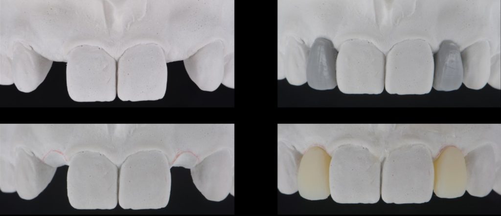
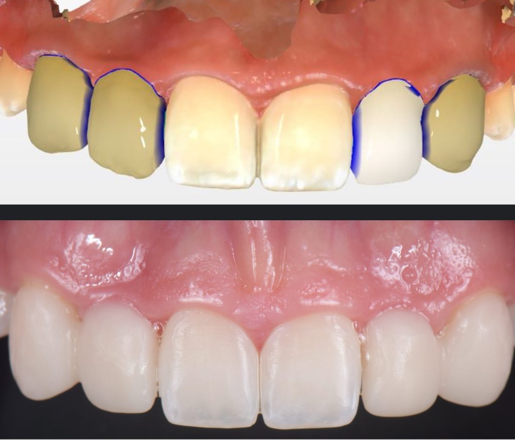
So, I needed to make sure that there was enough space for the restorative material. A purely additive wax-up was required to attain a mockup, verifying that the proposed contours were esthetically pleasing. This can be obtained either by an analogic approach (Fig. 15) or a digital approach (Fig. 16).
Incisally, there was no reduction required, the reduction was minimal, and from a cervical perspective, a 0.5 mm finish line was prepared (Fig. 17). This allowed our team to do a fundamentally additive design.
It is important here to emphasize that even if the age and preference of the patient allowed us to plan for an implant-supported restoration to replace the lateral incisors, the need to enhance the contours of the canines would still be required to provide a functional and esthetically pleasing result.
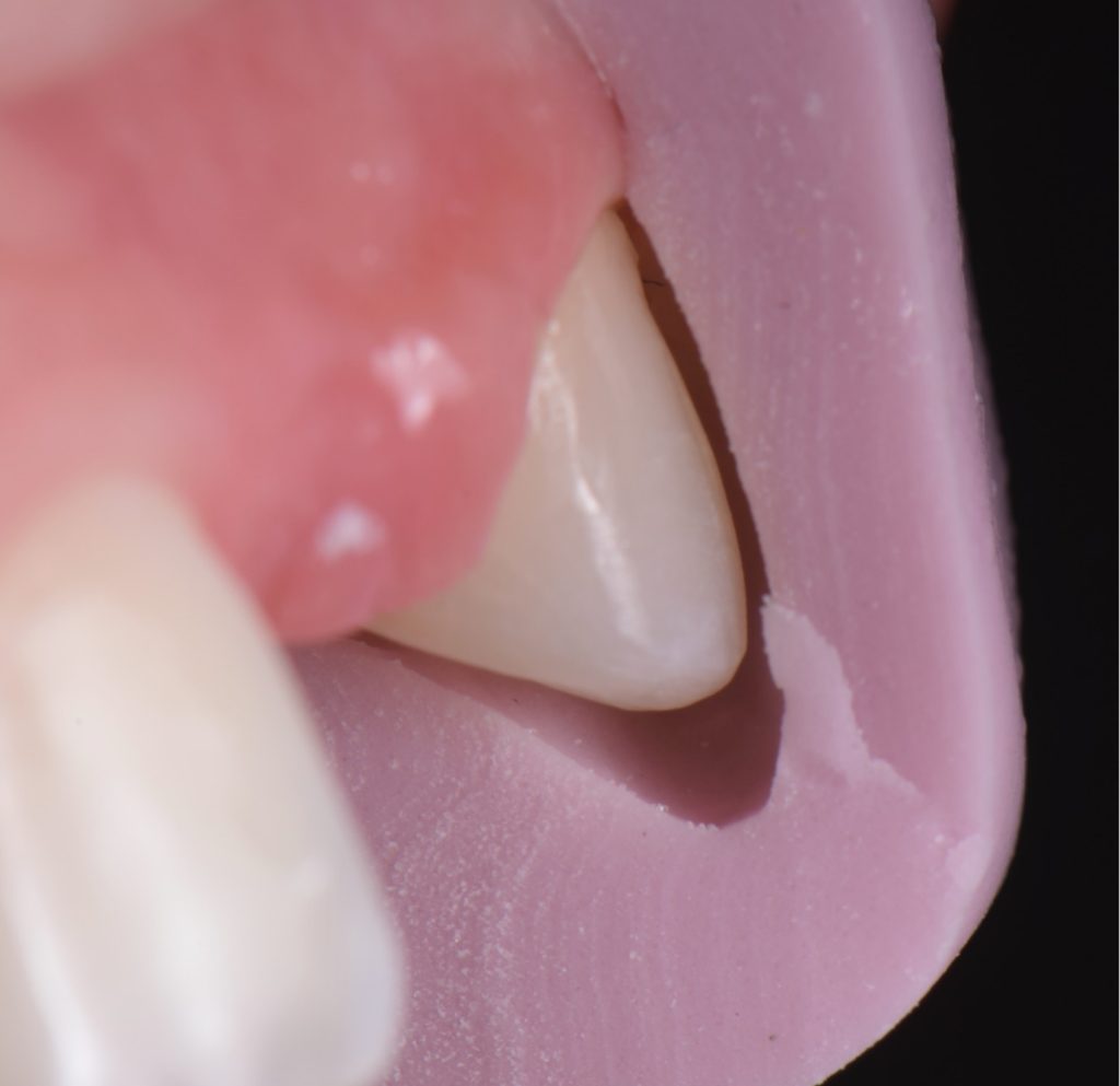
The undersized canines were out of function, removing all protective coverage for the lateral incisors. This provided a unique opportunity and indication for the most conservative scenario of a cantilevered RBFDP.
When considering a zirconia crown, we needed to ensure there was sufficient room for the wall thickness to be a minimum of 0.3 mm (ideally between 1.0 mm and 1.5 mm), an incisal reduction of 2.0 mm, and a visible and continuous circumferential chamfer with a reduction of at least 0.5 mm at the gingival margin. The patient’s preliminary condition allowed us to accomplish these space requirements with hardly any tooth reduction (Fig. 17).
RBFPD has an exceptional record, so this cantilever bridge can be considered a long-term solution. However, if in the future an implant-supported restoration is desired, the pontic of this cantilever can be removed, and an implant could be placed and restored.

Solutions for Congenitally Missing Lateral Incisors
This photo essay covers the first three solutions for addressing congenitally missing lateral incisors: canine substitution, resin-bonded single retainer bridge (resin-bonded fixed dental prostheses=RBFDP), and cantilever bridge. The next article in this discussion will address the fourth solution, a single-tooth implant.
SPEAR ONLINE
Team Training to Empower Every Role
Spear Online encourages team alignment with role-specific CE video lessons and other resources that enable office managers, assistants and everyone in your practice to understand how they contribute to better patient care.

By: Ricardo Mitrani
Date: June 21, 2022
Featured Digest articles
Insights and advice from Spear Faculty and industry experts
