The Straumann BLX implant system combines an innovative design with proven surface technology that gives the clinician the possibility of achieving high insertion torque during placement in a fresh extraction socket.

The concept of immediate implant placement after extraction provides reduction in the number of surgical interventions and a shorter treatment time. One of the shortcomings of this technique is that the implant diameter is often smaller than the diameter of the root of the extracted tooth, which may lead to a gap between the implant and the extraction socket. There are different protocols described in the literature to graft this area.

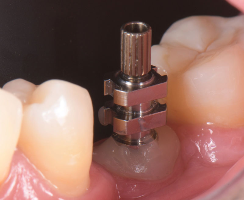
A simple protocol is described in this visual essay to walk you through the insertion of an immediate implant in a mandibular molar site in conjunction with a simple technique for grafting at the time of implant placement.

Patient presents with a fractured mandibular first molar with recurrent decay.
Periapical radiograph of the patient's mandibular first molar.
CBCT view of the site taken to analyze the amount of apical bone available above the inferior alveolar nerve canal.
The extraction was performed with minimal trauma to the hard and soft tissues maintaining the inter-radicular bone
A BLX WB 6.5x8 mm was inserted with 60 N/cm2. The implant was placed in position for a future screw-retained crown.
Periapical radiograph of the implant after placement.
Once the implant was placed, the surrounding area of the socket was filled with Bio-Oss collagen.
A collagen sponge was placed above the bone graft and resorbable synthetic sutures were placed to maintain the graft in place.
Periapical radiograph of the implant placed after healing abutment and bone graft.
Occlusal view after three months of healing with healing abutment.
Occlusal view after three months of healing without the healing abutment. Note the buccal contour.
A provisional screw-retained restoration was fabricated with an adequate buccal contour to support the soft tissue.
A provisional screw-retained restoration was inserted, and four weeks were given to allow sufficient time for soft tissue grooming.
Occlusal view after soft tissue grooming. Note the buccal contour.
Personalized impression coping was fabricated with composite.
Occlusal view of personalized impression coping assuring correct soft-tissue support.
Buccal view of personalized impression coping assuring correct soft-tissue support.
Periapical radiograph confirms the correct position of the impression coping.
Final VPS impression. Note how the personalized impression coping adequately transfers the submucosal contours.
Frontal view of the final zirconia crown bonded to a Variobase.
Occlusal view of the final restoration with the access hole sealed with composite. Note that the interproximal contact was optimized utilizing an inlay on the second molar.
Periapical radiograph 12 weeks post-insertion, with adequate bone maintenance around the implant.

Ricardo Mitrani, D.D.S., M.S.D., is a member of Spear Resident Faculty.