Beyond Titanium: The Revolution of Zirconia Dental Implants
By Joan Pi-Anfruns on June 22, 2020 | commentsThis article was co-authored by Yair Whiteman, D.M.D.
“I don't want any metals in my body.”
I can't tell you how many times I've heard this statement from patients, and I'm hearing it more and more. Patients are actively seeking alternatives to metals and data shows zirconia may be a viable option.
So, what's the issue with titanium?
Titanium has long been considered the material of choice and the standard of care for tooth replacement, with success and survival rates well above 90% after 10 years.1, 2
Despite the excellent results and biocompatibility reported for titanium implants, the demand for metal-free alternatives has increased. Patients are seeking alternatives to titanium for two main reasons:
- The belief they suffer from a metal allergy.
- They are seeking a holistic approach to their treatment (also referred to as biologic dentistry).
While the incidence of titanium allergy is low (0.6%), it can provoke type I and IV hypersensitivity reactions that resolve only with the removal of the implant.3 Additionally, titanium corrosion and particle release has been linked to changes in the DNA of peri-implant microorganisms, increased risk and incidence of inflammatory disease and peri-implantitis.4
In November 2019, a Federal Drug Administration (FDA) Advisory Panel meeting5 gathered a group of experts to review the evidence on immune system responses to metals and their clinical manifestations. Some of these manifestations include headache, vertigo, vision changes, fatigue, rash, joint and muscle pain, and weakness, among others.
According to the same report, more than 2.2 million adverse events related to dental implants have been reported to the FDA, making dental implants the second most implicated category of devices after hip prosthesis. The report also addresses patient susceptibility concerns and suggests a diagnostic or screening tool be developed to identify those patients who could potentially develop some of the manifestations.
With that, titanium dental implants have been, and continue to be, a reliable and successful option for tooth replacement. For many decades, millions of patients have benefited from this remarkable discovery. For many, it not only solves a simple problem of a missing tooth but instead, a life-changing event that allows patients to regain function, esthetics, self-esteem, and peace of mind.
The new kid on the block
Zirconia (zirconium dioxide, ZrO2)1 is a ceramic obtained from a reduction-chlorination reaction of zirconium, a metal. At the end of this process, zirconium dioxide powder (ZrO2)2 is obtained. This material is now a ceramic without any metallic properties of the raw material. It is both an electric and thermal insulator and resistant to corrosion.
Zirconia has been shown to have equal capacity for osseointegration when compared to titanium6, and ability to withstand occlusal forces.7,8 Investigation periods of up to five years have reported survival and success rates of more than 95% in humans9-11, making them a viable alternative for tooth replacement.
Commercially available zirconia implants can be one- or two-piece. One-piece zirconia implants with an integrated abutment and predetermined restorative margin have limitations due to their inability to allow for angle correction, require a high degree of surgical precision, and only accept a cementable restoration.
Advances in manufacturing have allowed for the development of two-piece systems with either cement or screw-retained abutments. These systems have allowed clinicians to broaden their prosthetic options, including angle correction via angled abutments, the ability to deliver screw-retained restorations as well as improved options for soft tissue management.
Patient demand for metal-free alternatives have increased significantly in the past five years and expected trends in global ceramic implant market shares predict that zirconia implants will capture about 10% of the market by 2025.12

Case report
Diagnosis:
A 34-year-old male with a noncontributory medical history presented with a hopeless tooth #9 due to endodontic failure. The patient had undergone previous orthodontic treatment, leading to root resorption and root canal therapy. The tooth was symptomatic, presented with Grade II mobility, and was treatment planned for extraction.
The patient was presented with two options to replace his missing tooth:
- A fixed partial denture prosthesis.
- A dental implant.
The patient elected to have the tooth replaced with a dental implant. He further requested a zirconia implant due to concerns with titanium particle release.

Treatment plan:
The case was treatment planned for extraction of tooth #9, immediate implant placement, and immediate provisionalization.
A CBCT (Accuitomo, J Morita USA; Irvine, California) was obtained to assess the site and in preparation for a digital workflow approach. Although the coronal 1/3 of the buccal plate was intact, the CBCT analysis revealed a fenestration on the facial apical 1/2 of the tooth.
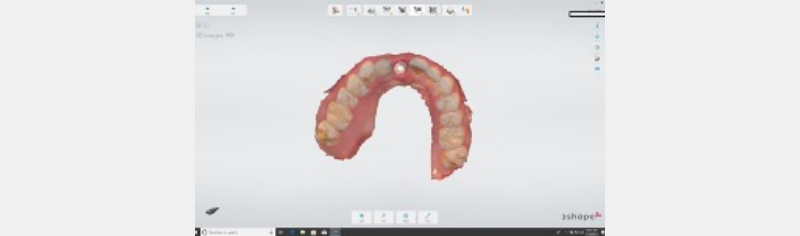
The DICOM files were uploaded to a digital planning software (CoDiagnostix®, Dental Wings GmbH; Chemnitz, Germany) and the case was planned for guided surgery. An intraoral scan was obtained prior to the extraction (Trios 3, 3Shape A/S; Copenhagen, Denmark) and the existing tooth was used as a reference in the planning software for proper positioning of the implant.
A PMMA provisional was designed and fabricated in preparation for the immediate provisionalization. A digital surgical guide was also generated and produced in a desktop 3D printer (Form 2, Formlabs; Somerville, Massachusetts).

Surgery:
After removal of the tooth and debridement of the socket, the surgical guide was seated, and the drilling protocol was followed per the manufacturer's recommendations following a guided protocol. A 4.1 x 12 mm RD PURE 2-piece Zirconia implant (Straumann®; Basel, Switzerland) was placed with an insertion torque of 35Ncm. The PMMA provisional shell was picked up intra-orally with flowable composite utilizing a customized Vita-CAD Temp® and shaped and polished extra-orally.
A tunnel approach was utilized and a resorbable collagen membrane was inserted. The site was grafted utilizing a bone graft complex consistent of PRF and Xenograft. Next, the PMMA provisional was primed using a universal adhesive primer (All-Bond Universal Bisco) and picked up intra-orally.
The provisional restoration was designed with palatal orientation wings, which were seated in intimate contact with the palatal aspect of the adjacent teeth to ensure ideal positioning of the restoration. The orientation wings were removed, and the provisional restoration was shaped, polished, and delivered.


Restoration:
Twelve weeks after implant placement, a final impression was obtained via intraoral scan and analogue methods with a custom impression coping. A zirconia framework with facial cutback was designed using the 3Shape Dental Manager software, duplicating the provisional restoration's transmucosal profile, as well as the coronal aspects, to fabricate a screw-retained layered zirconia restoration on the Straumann PUREbase abutment.
The restoration was tried in for contacts, occlusion, shape, and shade then prepared for final cementation and delivery. To ensure an accurate and reliable connection between the PUREbase and the zirconia restoration, an MDP-containing resin cement (Panavia V5, Kuraray America Inc.; New York) was utilized. The restoration was then torqued to 35Ncm and the screw access hole was plugged with Teflon tape and sealed with composite.


Joan Pi-Anfruns, D.M.D., is an assistant clinical professor at UCLA School of Dentistry, where he completed his Surgical Implant Fellowship in 2014. He lectures internationally on the topics of dental implants and bone regeneration.
Yair Whiteman, D.M.D., is the Director of the UCLA Center for Esthetic Dentistry and is an Assistant Clinical Professor in the Section of Restorative Dentistry in UCLA School of Dentistry. He received his dental degree from Semmelweis University, Budapest, Hungary, and a specialty certificate in prosthodontics at Boston University.
References
- Fischer K, Stenberg T. Prospective 10-year cohort study based on a randomized controlled trial (RCT) on implant-supported full-arch maxillary prostheses. Part 1: Sandblasted and acid-etched implants and mucosal tissue. Clinically Implant Dentistry and Related Research. 2012;14:808-815.
- Buser D, Janner SF, Wittneben JG, Bragger U, Ramseier CA, Salvi GE. 10-year survival and success rates of 511 titanium implants with a sandblasted and acid-etched surface: A retrospective study in 303 partially edentulous patients. Clinically Implant Dentistry and Related Research. 2012;14:839-851.
- Sicilia A, Cuesta S, Coma G, Arregui I, Guisasola C, Ruiz E, Maestro A. Titanium allergy in dental implant patients: a clinical study on 1500 consecutive patients. Clinical Oral Implants Research. 2008 Aug;19(8):823-35.
- Noumbissi S, Scarano A, Gupta S. A Literature Review Study on Atomic Ions Dissolution of Titanium and Its Alloys in Implant Dentistry. 2019 Jan 24;12(3):368. doi: 10.3390/ma12030368.
- https://www.fda.gov/media/131150/download
- Gahlert M, Rohling S, Wieland M, Sprecher CM, Kniha H, Milz S. Osseointegration of zirconia and titanium dental implants: a histological and histomorphometrical study in the maxilla of pigs. Clinical Oral Implants Research. 2009 Nov;20(11):1247-53.
- Andreiotelli M, Kohal RJ. Fracture strength of zirconia implants after artificial aging. Clinically Implant Dentistry and Related Research. 2009 Jun;11(2):158-66.
- Silva NR, Coelho PG, Fernandes CA, Navarro JM, Dias RA, Thompson VP. Reliability of one-piece ceramic implant. Journal of Biomedical Materials Research Part B: Applied Biomaterials. 2009 Feb;88(2):419-26.
- Grassi FR, Capogreco M, Consonni D, Bilardi G, Buti J, Kalemaj Z. Immediate occlusal loading of one-piece zirconia implants: five-year radiographic and clinical evaluation. International Journal of Oral Maxillofacial Implants. 2015 May-Jun;30(3):671-80.
- Oliva J, Oliva X, Oliva JD. Five-year success rate of 831 consecutively placed Zirconia dental implants in humans: a comparison of three different rough surfaces. International Journal of Oral Maxillofacial Implants. 2010 Mar-Apr;25(2):336-44.
- Balmer M, Spies B, Kohal RJ, Hammerle C, Vach, Jung R. Zirconia implants restored with single crowns or fixed dental prostheses: 5-year results of a prospective cohort investigation. Clinical Oral Implants Research. 2020 Jan 25. doi: 10.1111/clr.13581. [Epub ahead of print]
- Source: Vontobel Equity Research.