- Article
- Occlusion / Wear
Vertical Dimension in Dentistry: Dentate and Edentulous
Greggory Kinzer
•
July 9, 2018
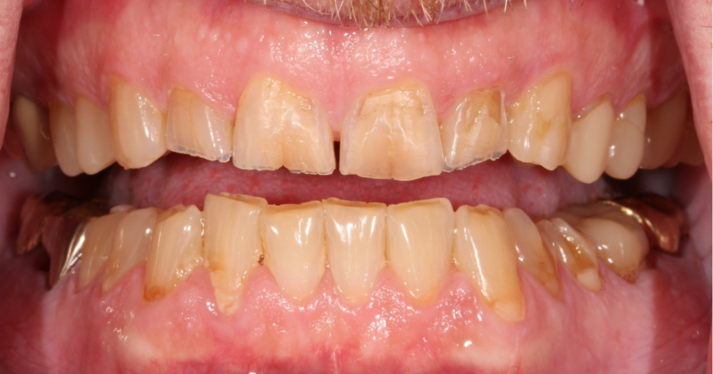
Vertical dimension in dentistry refers to the distance between the maxilla and mandible when a patient’s teeth are in contact, and it plays a central role in nearly every restorative and prosthodontic treatment plan.
The concept of using rest position to determine the correct vertical dimension when restoring patients typically works well for edentulous patients, but has limitations for dentate patients.
Because the diagnostic challenge varies depending on whether a patient has natural teeth, this guide addresses vertical dimension determination for dentate and edentulous patients separately, along with the criteria that help clinicians determine when a change in vertical dimension is warranted.
The following will discuss some philosophies that can be used to determine the correct VDO in patients who still have their natural teeth.
Determining vertical dimension in dentate patients
Trial appliance
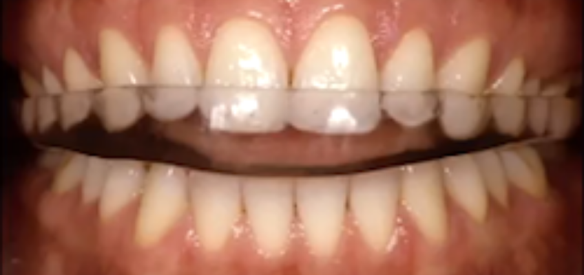
With this protocol, a patient is typically asked to wear an acrylic appliance for three months to evaluate whether the desired vertical dimension can be tolerated. The rationale for this method is that the patient will experience pain if the vertical dimension is unacceptable.
However, except in a few patients with temporomandibular joint problems, altering vertical dimension does not produce pain. Although the appliance may be very useful for determining other elements of treatment or for aiding muscle deprogramming, it does not provide specific information regarding vertical dimension.
Measurements using the cementoenamel junction
Another method to determine vertical dimension is to measure from the cementoenamel junction (CEJ) or gingival margin of the maxillary central incisors to the CEJ or gingival margin of the mandibular central incisors.
This distance is then compared to the 18–20 mm average distance seen in a dentition of unworn teeth and a Class I occlusion. If this distance is less than 18 mm, it likely indicates a loss of vertical dimension and therefore supports increasing the VDO.
The primary flaw in this approach is that the anterior teeth don’t establish the VDO; the length of the ramus and the eruption of the posterior teeth establish it. Measuring the distance between the CEJ or gingival margins merely represents the amount of anterior tooth eruption, not the vertical dimension of occlusion. Indeed, it’s possible to have an extremely diminished CEJ-to-CEJ distance in the anterior and a perfectly normal vertical dimension of occlusion.
This situation commonly occurs in patients with severe anterior tooth wear and no posterior tooth wear. Most clinicians examine the worn anterior teeth and decide to open the bite to gain space for restoration, when in fact the patient could be treated at the existing vertical dimension by intruding the worn anterior teeth or crown-lengthening them to correct the gingival levels.
As a general rule, it’s highly unlikely that a patient has lost vertical dimension if the posterior teeth are present, unworn, and in occlusion. If space to restore the anterior teeth is lacking, it’s also likely that orthodontics or crown lengthening would allow the patient to be treated without the need to treat their posterior teeth.
Managing patients with severe tooth wear often requires balancing restorative space, occlusion, and long-term function. Dentists who want to deepen their approach to these cases can continue their learning in Spear’s Worn Dentition workshop.
Transcutaneous electrical neural stimulation
A third method for determining vertical dimension, used for decades, is transcutaneous electrical nerve stimulation.
With this approach, electrodes are placed over the coronoid notch, and a mild, cyclic electrical current is applied to stimulate contraction of the masticatory muscles via the cranial nerves. The surface electrical activity of the temporalis, masseter, and digastric muscles is recorded electromyographically, and a jaw-tracking device evaluates the position of the mandible relative to the maxilla.
A baseline electromyographic reading is taken before any muscle relaxation. The TENS unit is then programmed to relax the masticatory muscles, and the electrical activity of the muscles is again evaluated.
Neuromuscular rest is achieved when the elevator muscles are at their lowest level of activity, with no increase in the electrical activity of the digastric muscles. This neuromuscular rest position is thought to be the starting point for building occlusion. The operator closes up from this position for the “new” amount of freeway space, effectively using the combination of neuromuscular rest and freeway space to determine the new occlusal vertical dimension.
The primary flaws in this approach relate to the neuromuscular adaptability of patients. The resting electrical activity of muscles, like the freeway space, returns to pretreatment levels within 1–4 months after treatment.
Moreover, this approach often results in a more open vertical dimension than the patient’s existing one, which can necessitate extensive restorative dentistry and extremely large teeth to accommodate the vertical dimension dictated by the TENS device.
When to change vertical dimension: 5 decision criteria
Measuring or estimating vertical dimension is only part of the equation. Before altering the VDO of a worn dentition, clinicians should weigh five criteria to determine whether a change is actually warranted: prosthetic height, anterior occlusal relationship, skeletal typology, TMJ health, and facial esthetics.
#1 Prosthetic height
One of the primary factors in determining whether a vertical dimension change is needed is the amount of tooth structure available for restoration. If adequate tooth structure remains, no change in vertical dimension may be necessary. If restorative space is lacking, opening the vertical dimension becomes appropriate.
The literature references a minimum of 4 mm of posterior tooth preparation height for resistance and retention form. However, modern materials and adhesive systems may push that limit to 3 mm, provided the ferrule is adequate. Crown-lengthening surgery can help when crown height falls below 3 mm, but the remaining crown-to-root ratio must also be considered.
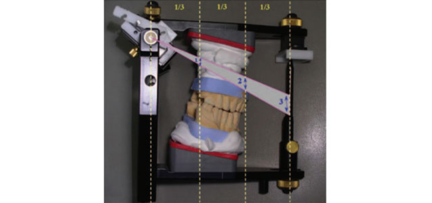
The “Rule of Thirds” is a useful clinical reference: roughly 1 mm of vertical opening in the posterior produces about 3 mm of corresponding opening in the anterior. This ratio is easily visualized on an articulator and can be used from a centric relation position to gain anterior restorative space with minimal posterior change.
Applying occlusal principles consistently across restorative cases takes both a solid diagnostic process and clinical repetition. Dentists interested in refining these skills can explore Spear’s Occlusion in Everyday Practice: Managing Patients with Simple to Complex Needs seminar.
#2 Anterior occlusal relationship
The anterior tooth relationship relates to overbite and overjet. In a normal Angle Class I incisor relationship, overjet averages 3–4 mm and overbite averages 2–3 mm. These functional contacts are important for anterior guidance and must be maintained.
When the overbite is too deep (greater than 4 mm) and the overjet is too minimal (less than 2 mm), tooth interference may lead to pathway wear, tooth mobility, interproximal opening, and tissue irritation.
Too little overbite and overjet can result in a lack of anterior guidance and increased posterior wear. Changing vertical dimension can help resolve either situation by improving function, reducing wear patterns, and minimizing the amount of tooth preparation necessary.
In an open bite situation, closing the vertical dimension brings the anterior teeth closer together, creating a more ideal relationship. Opening a deep bite increases the distance between the incisors and provides shallower guidance, decreasing overbite and overjet.
Care must be taken with Class II situations, since downward rotation of the mandible can increase overjet. Facebow-mounted models help visualize these changes as VDO is opened or closed.
#3 Skeletal typology
Mandibles can grow in either vertical or horizontal directions, producing facial growth patterns classified as dolichofacial (long-face, hyperdivergent), brachyfacial (short-face, hypodivergent), and mesofacial (medium- or neutral-face, normodivergent).
Ramus height and gonial angle are key determinants of skeletal facial type. An acute gonial angle is associated with a flat mandibular plane and a brachyfacial type, while a dolichofacial type has an obtuse gonial angle and a steeper mandibular plane. VDO is the result of musculoskeletal balance during growth, so cephalometric analysis does not provide an ideal value for vertical dimension on its own; however, the skeletal pattern can indicate how predictable opening or closing of the OVD is likely to be.
#4 TMJ
A stable TMJ is crucial for a successful change in occlusal vertical dimension, since strict rotation around the hinge axis is used as the reference point for VDO determination. Centric relation, or an adapted centric posture, must be attainable before proceeding.
Intra-articular disease or dysfunction must be evaluated and stabilized first. A complete TMJ examination looks for disc displacement, crepitus, mandibular mobility, and the joint’s ability to be loaded under function, supplemented by radiographic analysis (panoramic X-ray, CBCT, or MRI as needed).
Evidence of osteoarthritic change or condylar surface abnormality precludes a vertical dimension change until stabilization occurs. Patients who may have difficulty adapting to neuromuscular change, such as those with Parkinson’s disease or some elderly patients, should generally be avoided as candidates. Occlusal splint therapy can help stabilize and evaluate joint position before comprehensive restorative treatment.
#5 Facial esthetics in dentate patients
The determinants of facial esthetics are the sagittal profile, facial tissue appearance, lip morphology, and tooth display. A pleasant facial height is the target for a change in vertical dimension; changing VDO will not affect the upper lip dynamic relative to tooth display at rest unless a maxillary osteotomy is involved.
In normal anatomical facial analysis, mid-face and lower-face heights should be approximately 50/50 relative to each other. Loss of VDO due to tooth wear can negatively affect facial appearance, including pseudo-prognathism from forward mandibular rotation, altered facial contour, narrowed vermillion borders, and an over-closed commissure.
That said, increasing VDO through restoration, even by as much as 6 mm, may not change facial appearance on its own, and increasing VDO should not be undertaken solely to improve facial esthetics.
Although facial appearance, TMJ health, and skeletal typology are important parameters, the primary motivators for a change in VDO remain restorative space requirements and anterior tooth relationships.
For clinicians looking to strengthen their diagnostic approach to occlusal vertical dimension, centric relation, and treatment planning, Spear’s Occlusion in Clinical Practice workshop provides hands-on instruction for applying these principles in everyday restorative care.
Determining vertical dimension in edentulous patients
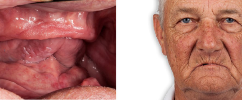
Vertical dimension in dentistry looks markedly different once a patient has lost all their natural teeth.
When an edentulous patient presents for restorative treatment, determining the optimal occlusal vertical dimension is one of the most common clinical challenges, particularly if the patient presents without existing prostheses to reference. Both the literature and clinical experience offer a range of preextraction and postextraction options for making this determination.
If a patient presents with existing conventional complete dentures, a common approach is to transfer that vertical dimension to the newly fabricated prostheses. This can be useful information, but it is potentially risky because it assumes the existing OVD is correct, which may not be true.
Pre-extraction vs. post-extraction methods
Occlusal vertical dimension is defined as the distance between two selected anatomic or marked points when in maximal intercuspal position. Clinically, the two points most commonly used are the tip of the nose and the tip of the chin; because these points are arbitrary, the absolute distance between them is not itself a valuable indicator of OVD.
Pre-extraction records, when available, can be useful for determining OVD and include intraoral measurements, soft-tissue profile tracings, cephalometric measurements, and photographs. Relying solely on pre-extraction records has two limitations: the records are often unavailable, and even when available, they assume the pre-extraction vertical dimension was correct, which may not be the case. As a result, a post-extraction method is frequently required.
Post-extraction methods for OVD determination include freeway space, following existing dentures, facial esthetics, phonetics, swallowing, craniofacial landmarks, cephalometric evaluation, and several additional methods. The most widely used post-extraction method is the freeway space approach.
Freeway space approach
Physiologic rest position, or rest vertical dimension, is the postural position of the mandible when a person is resting comfortably, and the associated muscles are in minimal contractile activity. Freeway space, or interocclusal rest distance, is the difference between the rest vertical dimension and the occlusal vertical dimension.
With this method, the physiologic rest position is identified clinically, and an average freeway space value (2–4 mm) is subtracted to arrive at the proposed occlusal vertical dimension.
Common ways to identify the physiologic rest position include asking the patient to relax with the lips together, to lick their lips, swallow, and relax, or to make an “em” sound and evaluate the reference points as the sound finishes. In practice, more than one of these techniques is often needed.
The main limitation of this approach is the repeatability of the physiologic rest position. The literature is mixed on whether this position is stable over time, so it is often more accurate to think of physiologic rest as a range rather than a single, reliably repeatable position. This technique has a strong clinical track record as a starting point, but clinicians should not rely on it alone.
Following the existing dentures
When a patient presents with dentures, a common approach combines the existing OVD with the freeway space approach. With the dentures inserted, have the patient relax and evaluate the interocclusal distance between the maxillary and mandibular premolars. A space of less than 2 mm suggests the OVD may be excessive; greater than 4 mm suggests it may be deficient; a measurement between 2 and 4 mm is generally considered correct.
The risk with this approach is assuming the existing dentures’ OVD is correct. In clinical practice, denture teeth wear, and supporting tissues resorb over time, typically resulting in a deficient OVD in an existing set of dentures. Patients also show variability in physiologic rest position depending on whether dentures are in place during evaluation. As a result, existing dentures are best used in combination with the freeway space approach rather than followed on their own.
Facial esthetics for edentulous patients
As with dentate patients, an ideal OVD achieves a normal facial proportion of roughly 50% of the lower one-third to 50% of the middle one-third of the face. A common finding in denture wearers is that the lower one-third of the face is deficient relative to the middle one-third, indicating an OVD deficiency.
Phonetics
The phonetic approach extends the physiologic rest concept using the /M/ sound and subtracting 2–4 mm, and also uses the /O/ and /E/ sounds.
One study describes asking patients to pronounce /O/ and subtracting 5.5 mm, and /E/ and subtracting 7.5 mm, to establish the desired OVD. Challenges include variability in vowel pronunciation across languages and the practical difficulty of measuring distances while a patient is speaking.
Despite these challenges, phonetics is an efficient way to validate an OVD established with another technique.
Swallowing
The swallowing approach relies on the mandible moving from the physiologic rest position to tooth contact during swallowing. The classic technique uses soft wax on record bases while the patient swallows.
Due to the difficulty of managing the wax and variability in swallowing duration and intensity, this approach is generally considered better suited to verifying an OVD than establishing it initially.
Craniofacial landmarks
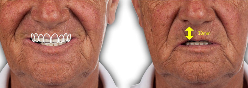
This approach uses facial landmarks and measurements between them to approximate the desired height of the lower one-third of the face, and therefore the desired OVD.
For example, maxillary incisal edge position is determined by desired tooth display and upper lip length, lower incisal edge position by desired tooth display relative to the lower lip, and OVD by the desired vertical overlap of the anterior teeth.
This approach offers strong esthetic predictability, but most clinicians don’t set denture teeth themselves. They may find it difficult to communicate the desired OVD to a laboratory technician, making it more straightforward for natural teeth than for edentulous patients.
Cephalometric evaluation
Cephalometric landmarks are useful for OVD determination because specific landmarks do not change following edentulation. Practical constraints have historically limited this approach, though low-exposure CBCT protocols and virtual articulation are making radiographic landmarks increasingly relevant to predictable OVD determination.
Additional methods: the tactile approach
Several additional methods exist for determining OVD, ranging from finger-length correlations to bite force capacity. One approach worth highlighting is the tactile approach, which uses a device with an adjustable jackscrew, such as an intraoral tracer, to gradually alter OVD based on direct patient feedback about whether the bite feels too “tall” or too “short.” The key advantage of this approach is that it engages the patient directly in evaluating and determining the desired OVD.
Choosing the best approach for edentulous patients
Determining vertical dimension is only one component of successful full arch rehabilitation. Spear’s Restoring the Edentulous Arch workshop explores the complete restorative workflow, from diagnosis and treatment planning through predictable prosthetic execution.
No single method described here works for every edentulous patient every time, so familiarity with multiple approaches builds confidence in managing these cases.
For many patients, establishing vertical dimension in dentistry begins with the freeway space and existing denture methods, is then confirmed through facial esthetics, phonetics, and swallowing, and is finally refined with the tactile approach.
If the proposed occlusal vertical dimension doesn’t feel right to the patient, it’s better to make adjustments at the wax rim or trial denture stage than after the prosthesis reaches final fabrication. Vertical dimension is one of several factors that determine restorative success. See Spear’s guide to occlusion fundamentals for the complete framework
Contributing Authors
Dr. Jeffrey Bonk & Dr. Darin Dichter
References
- Abduo, J., & Lyons, K. (2012). Clinical considerations for increasing occlusal vertical dimension: a review. Australian Dental Journal, 57(1), 2-10.
- Ahila, S. C., Sasikala, C., Kumar, B. M., Rajdeep, T., & Abinaya, K. (2016). Evaluation of the correlation of ramus height, gonial angle, and dental height with different facial forms in individuals with deep bite disorders. Annals of Medical and Health Sciences Research, 6(4), 232-238.
- Alhajj, M. N., Khalifa, N., Abduo, J., Amran, A. G., & Ismail, I. A. (2017). Determination of occlusal vertical dimension for complete dentures patients: an updated review. Journal of Oral Rehabilitation, 44(11), 896-907.
- Igić, M., Krunić, N., Aleksov, L., Kostić, M., Igić, A., Petrović, M. B., et al. (2015). Determination of vertical dimension of occlusion by using the phonetic vowel “O” and “E”. Vojnosanitetski Pregled, 72(2), 123-131.
- Orthlieb, J. D., Laurent, M., & Laplanche, O. (2000). Cephalometric estimation of vertical dimension of occlusion. Journal of Oral Rehabilitation, 27(9), 802-807.
- Pepicelli, A., Woods, M., & Briggs, C. (2005). The mandibular muscles and their importance in orthodontics: a contemporary review. American Journal of Orthodontics and Dentofacial Orthopedics, 128(6), 774-780.
- Rebibo, M., Darmouni, L., Jouvin, J., & Orthlieb, J. D. (2009). Vertical dimension of occlusion: the keys to decision. International Journal of Stomatology & Occlusion Medicine, 2(3), 147-159.
- Edition, N. (2017). The glossary of prosthodontic terms. The Journal of Prosthetic Dentistry, 117, e20.
- Turrell, A. J. (1972). Clinical assessment of vertical dimension. The Journal of Prosthetic Dentistry, 28(3), 238-246.
Frequently Asked Questions
SPEAR campus Workshops
Learn Hands-on From Faculty Who Still Practice
A few days at Spear Campus can change how you diagnose, plan, and present cases, not through more lectures, but through hands-on training led by faculty who treat patients every week. Work through real cases in small groups and leave with techniques you can use on your next patient.
Featured Digest articles
Insights and advice from Spear Faculty and industry experts