Diagnosing TMD: Similarities Between Spear’s Occlusal Exam and the DC/TMD
By Curt Ringhofer on June 3, 2025 |Ever since the American Dental Association recognized orofacial pain as the 12th dental specialty in 2020, there has been confusion about who can, when to, and how to treat temporomandibular joint disorders. This series of two articles clarifies the similarities between the Spear occlusal examination and the one adopted by orofacial pain specialists, known as the Diagnostic Criteria for Temporomandibular Disorders.
An occlusal exam with few false positives and negatives
Temporomandibular joint disorders are estimated to affect 5%–12% of the adult population. However, these numbers may be surprisingly low when factoring in asymptomatic patients who show signs of a mandibular growth disturbance.
The Research Diagnostic Criteria for Temporomandibular Disorders, published in 1992, was a protocol researchers and clinicians could use for diagnostic and treatment purposes. However, the RDC/TMD wasn’t intended to be a comprehensive or self-sufficient system for all temporomandibular disorders, so in 2012, it was superseded by the DC/TMD, which has a more comprehensive classification system with higher sensitivity and specificity.
A high-sensitivity exam more accurately identifies patients with a disease, resulting in low false positives.
An exam with high specificity, meanwhile, more accurately identifies people without a disease, leading to low false negatives.
The goal for the DC/TMD is to achieve a sensitivity ≥ 0.70 and a specificity ≥ 0.95.
How Spear’s exam stacks up against the DC/TMD
Like its predecessor, the DC/TMD uses Axis I and Axis II protocols.
Axis I, which involves the physical assessment, is used during new-patient examinations to diagnose and screen for TMJ disorders and establish a treatment protocol.
Axis II assesses the patient’s psychosocial status and pain-related disability.
The DC/TMD exam remains the standard for screening patients with such disorders, but a Spear-trained dentist will have already gathered all the required information for a tentative diagnosis during their regular patient examination. This includes recognizing structurally altered temporomandibular joints and when imaging is necessary for a diagnostic approach to treatment.
In 2024, the International Network for Orofacial Pain and Related Disorders Methodology established 10 key points that summarize the current understanding of the etiology, diagnosis, and treatment of TMD patients. I’ll use these points as talking points and compare the DC/TMD examination to those taught in Spear workshops.
Point 1: “Patient-centered decision-making alongside patient engagement and perspective is critical to manage TMDs, with management being the process from history through examination into diagnosis and then treatment. Expectations should focus on learning to control and manage the symptoms and decrease their impact on the individual’s everyday life.”
The Spear new-patient examination starts with the initial interview. This allows the dentist to understand the impact TMD has had on the patient and opens a conversation that will guide the direction of treatment.
It’s imperative that the patient, the dentist, and the team agree on a diagnosis before initiating treatment. The dentist must discuss this during the initial interview and set expectations before treatment starts so the dental team can align itself with the patient’s expected outcome.
In addition, many patients seeking treatment for TMJ disorders have visited multiple dentists before their first appointment with a Spear-trained dentist. The initial interview gives the dentist the information needed to understand the treatment already provided.
SEPT. 11–12 AT SPEAR CAMPUS IN SCOTTSDALE, ARIZONA Don’t Miss This 2-Day Occlusion Seminar! Can you safely use a patient’s existing occlusion for your treatment … and if not, then what? |
Point 2: “TMDs are a group of conditions that may cause signs and symptoms, such as orofacial pain and dysfunction of a musculoskeletal origin.”
Historically, TMD has been associated with pain, but pain is only one component of temporomandibular joint disorders. And although the DC/TMD recognizes that pain is a driving factor for patients to seek treatment, growth disturbances can also be a reason for treatment.
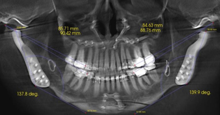
At Spear, we teach that a growth disturbance may be an early sign of a structurally altered temporomandibular joint, and a retrognathic mandible is a sign of a possible disc displacement. The condyle is one of the primary growth centers of the mandible, and a disc displacement can inhibit growth and development of the maxilla – mandibular complex.
Identifying and addressing growth disturbances early is critical because it allows the dentist to recommend treatment based on a diagnosis derived from imaging. A CBCT and an MRI are necessary to determine if growth is possible.
Figs. 1a and 1b illustrate a condyle with a favorable prognosis. The incomplete cortical plate formation and disk reduction in the Class I canine position provide information that enables the dentist to communicate the prognosis to the patient and their parents. A functional appliance could foster condylar growth, but if the disk doesn’t reduce in the treatment position of a functional appliance, this may lead to a more severe growth disturbance (Figs. 2a and 2b).


Point 3: “The etiology of TMDs is biopsychosocial and multifactorial.”
The etiology of occlusal disease is a primary focus of the examination process taught at Spear. As previously mentioned, TMD can manifest as pain or changes in occlusion. Either can originate from one or more of the following areas: muscle, disc, bone, cervical spine, or sympathetic hyperactivity.
Muscle pain (myalgia) can be the primary cause of the pain, but often it’s secondary to an underlying condition such as an airway disturbance, articular disc displacement, condylar breakdown, or cervical spine imbalance. It’s the responsibility of the treating dentist to determine the source of the muscle pain. When there’s an underlying reason for muscle pain, the muscles will be in hypercontraction “protection mode” to inhibit the mandible from moving in a direction that could lead to pain, such as the condyle pushing the disc into a muscle or a nerve.
Sympathetic hyperactivity can manifest as a condition known as complex regional pain syndrome, but it often appears as a sleep disturbance. During restful sleep, there is a transition from sympathetic to parasympathetic mode. Sympathetic activity is prevalent during wakefulness and REM sleep, while parasympathetic activity dominates during deep or Stage 3 sleep, essential for healing. A disruption in sleep quality may lead to sympathetic hyperactivity, potentially contributing to muscle dystonia and heightened pain perception.
The Spear curriculum addresses the biopsychological and multifactorial nature of TMDs and focuses on a diagnosis-first approach, as stated in the DC/TMD literature.
Point 4: “Diagnosis of TMDs is based on standardized and validated history taking and clinical assessment performed by a trained examiner and led by the patient perspective.”
The Spear curriculum, designed as a mini-prosthetic residency, aligns with the DC/TMD examination protocols. As previously mentioned, the new patient examination begins with an interview process, during which a detailed history is obtained, focusing on pain, trauma, joint clicking, joint locking, muscle pain, and airway disturbances.
Up next: Treatment protocols
This article focused on obtaining a diagnosis through clinical examination and how the DC/TMD and Spear clinical examinations gather similar data for a tentative diagnosis.
Part 2 of this series addresses the similarities between the DC/TMD and Spear treatment protocols for patients suffering from TMD. The difference is that the DC/TMD examination concerns pain, while the Spear occlusal examination adheres to the ADA-recognized evaluation of the temporomandibular joints and expands the assessment to include occlusal stability for the restorative dentist.
References
- Manfredini, D et al. (2024). Temporomandibular disorders: INfORM/IADR key points for good clinical practice based on standard of care. CRANIO, 43(1), 1–5. Link
- Schiffman, E et al., Orofacial Pain Special Interest Group, International Association for the Study of Pain (2014). Diagnostic criteria for temporomandibular disorders (DC/TMD) for clinical and research applications: Recommendations of the International RDC/TMD Consortium Network and Orofacial Pain Special Interest Group. Journal of Oral & Facial Pain and Headache, 28(1), 6–27. Link
- Piper M. (2020). Temporomandibular joint imaging. In R. Kerstein (Ed.), Handbook of Research on Clinical Applications of Computerized Occlusal Analysis in Dental Medicine (pp. 582–697). IGI Global Scientific Publishing. Link
- Nebbe B, Major PW & Prasad NG (1998). Adolescent female craniofacial morphology associated with advanced bilateral TMJ disc displacement. European Journal of Orthodontics, 20(6), 701–712. Link
- Choy EH (2015). The role of sleep in pain and fibromyalgia. Nature Reviews. Rheumatology, 11(9), 513–520. Link