- Article
- Implants
Photogrammetry for Dental Implants: The Cost of Efficiency
Leila Zadeh
•
September 22, 2026
Rethinking accuracy and responsibility in full-arch dentistry
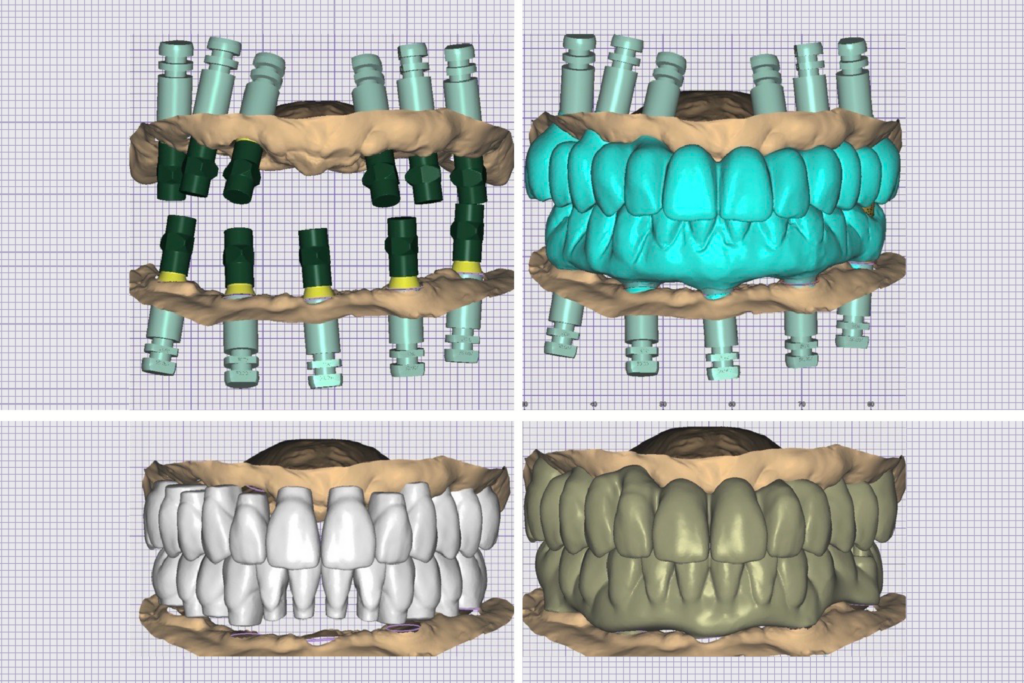
Success with full arch implant prostheses depends on understanding how chairside clinical choices, including the growing use of photogrammetry for dental implants, shape outcomes at the lab bench. As a dual-trained dentist and technician, I care deeply about the intersection of clinical dentistry and laboratory work, and I have learned that “good dentistry” in complex restorative cases is rarely achieved by any single tool.
Instead, it is the culmination of how accurately our records capture reality, and how faithfully they translate through a workflow, with as little distortion as possible, until the final prosthesis is seated and maintained over time.
Photogrammetry for dental implants, or PG, is one of the more powerful examples of this principle in modern implant dentistry, and some common examples on the market include the iCam4D (iMetric), MicronMapper (S.I.N. 360), PIC System (PIC Dental), and Blue Sky Bio Grammee (powered by Tupel 3D).
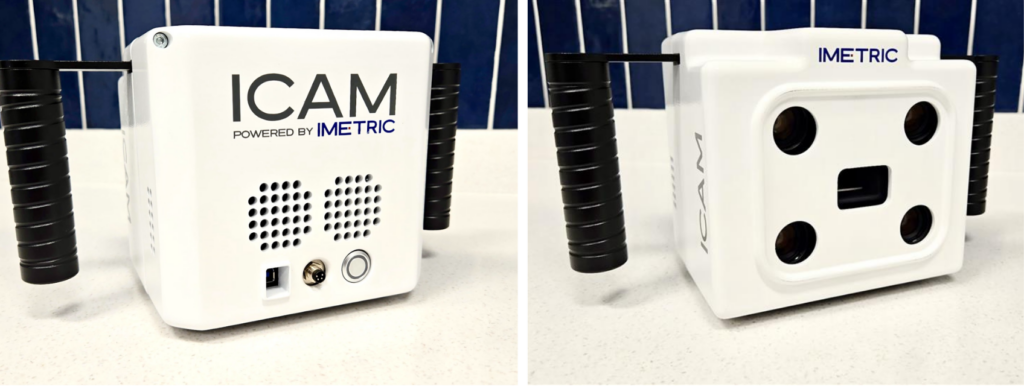
For fixed, multi-unit implant prostheses, photogrammetry for dental implants can help speed up the final impression appointment by reducing steps, and it can be a reliable tool for recording implant positions.4,5
However, PG is an expensive technology not without challenges and added responsibilities, especially when efficiency becomes the primary motivator rather than quality.
The challenge of full arch analog workflows
If you have ever restored a full arch fixed implant prosthesis using a traditional analog workflow, you know the feeling: an entire morning or afternoon block can be consumed, the patient becomes restless, and the operator takes on the roles of clinician, laboratory technician, therapist, and project manager simultaneously. Even when everything goes “well,” the process can be technique-sensitive and time-intensive.
The issue is not that analog methods are inherently inferior. The issue is how many steps they often require. And the more steps involved in any workflow, the higher the risk of cumulative error. Each step introduces variability, including material distortion, component seating discrepancies, resin shrinkage, cast inaccuracies, mounting errors, and operator technique. Even small deviations can become clinically meaningful when passive fit is compromised.
While digital workflows do not eliminate error, they can reduce the number of opportunities for error to be introduced. And photogrammetry for dental implants is a prime example.
What does photogrammetry for dental implants actually capture?
Photogrammetry for dental implants approximates three-dimensional relationships from two-dimensional images. In implant dentistry, PG systems capture the 3D spatial relationships between implants. That relationship can then be exported and used in computer-aided design (CAD) software to design a framework or fixed multi-unit implant prosthesis.1
Why is this important? Because passive fit is a requirement for long-term mechanical stability and biologic health. Inaccurate implant position transfer can introduce stress into the prosthesis-implant complex and increase the risk of complications, including screw loosening, fracture, and peri-implant bone loss over time.2,3
One reason photogrammetry for dental implants has gained traction is its ability to essentially bring the precision of a controlled extraoral scanner (i.e., a desktop scanner) to the mouth. This is especially important for long edentulous spans, where each additional implant increases the complexity of accurately transferring these spatial relationships and, therefore, increases the risk of prosthesis misfit.
How accurate is photogrammetry for dental implants across different full arch impression workflows?
It is tempting to assume that an intraoral scan alone can serve as the final impression for a full arch implant restoration.
In reality, intraoral scanning of edentulous arches remains challenging because these scanners perform best when capturing rigid, non-movable structures with unique surface detail, such as teeth. Intraoral scanners rely on linking multiple sequential images, and this stitching process is more likely to accumulate errors over long spans.
Additional factors such as scan path, tissue mobility, saliva and blood, poor-quality oral landmarks, and patient movement can further compromise accuracy.
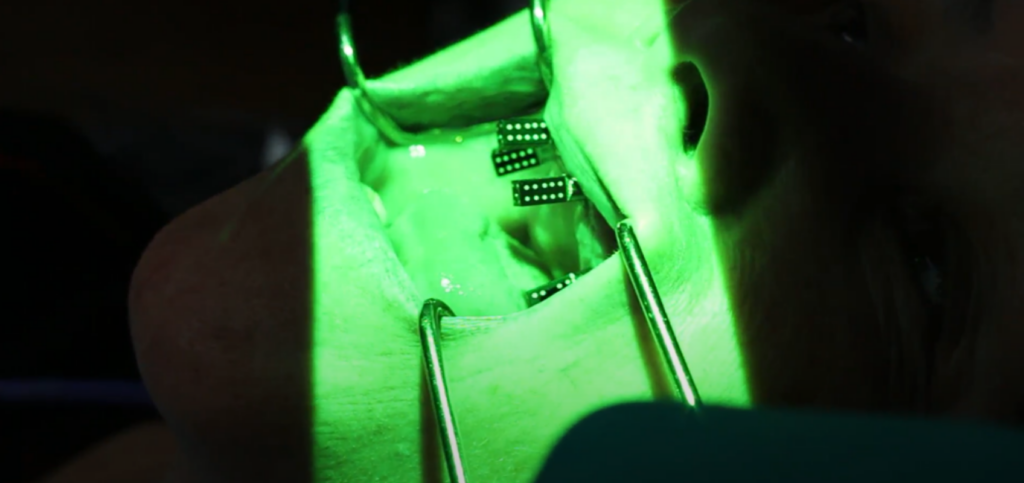
Photogrammetry for dental implants bypasses the issue of stitching error, which is why PG is positioned as a high-accuracy method for full arch implant position transfer. However, while photogrammetry for dental implants can improve the accuracy of capturing long-span implant spatial relationships when compared with intraoral scanning alone, it is not a perfect solution. PG accuracy is influenced by the specific system used, the condition, positioning, and seating of implant components and markers, and the clinical environment in which the data is captured. 1,3,4,6
When comparing photogrammetry for dental implants, intraoral scanning, and conventional analog workflows, it is essential to recognize that accuracy is influenced by how the impression process is performed. This is why meaningful comparisons require careful consideration of system design, component selection, clinical protocol, and operator experience. Continued research is needed to further investigate how these variables influence accuracy in full arch implant impressions.
Where does photogrammetry for dental implants add the most value?
Photogrammetry for dental implants is useful in situations involving long edentulous spans, which carry significant consequences for small inaccuracies due to strict passive-fit requirements.
In these cases, reducing the number of technique-sensitive impression steps to capture implant positions can improve efficiency and also simplify appointments that historically required substantial chair time.
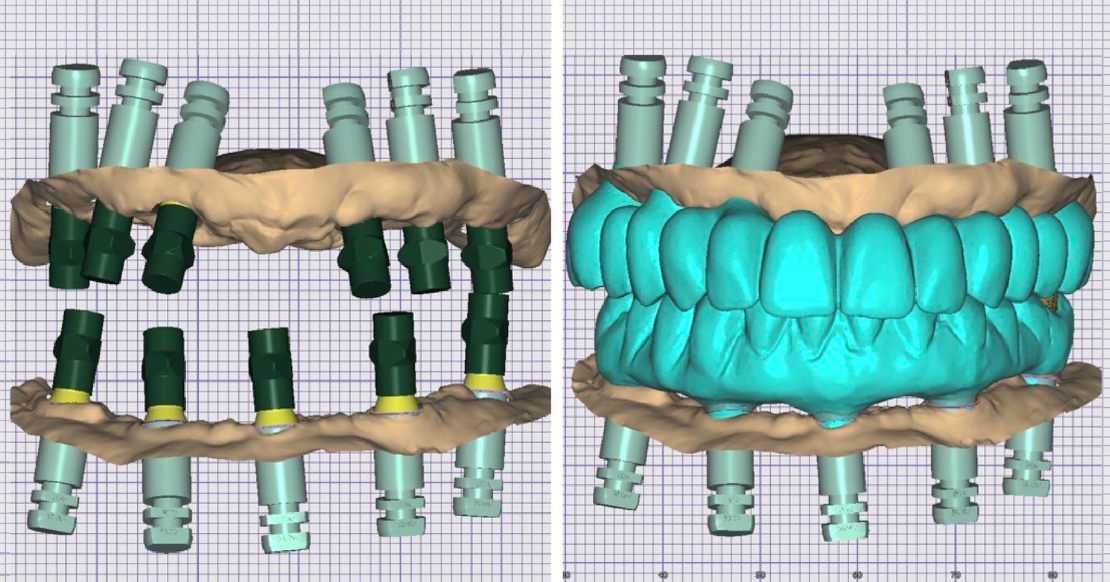
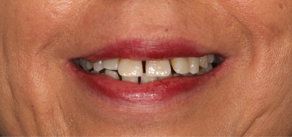
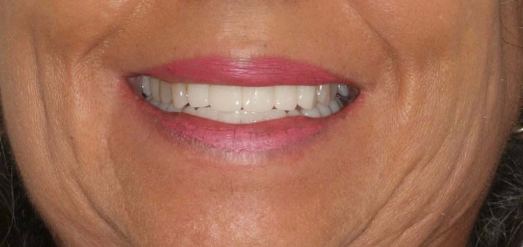
Although most commonly associated with full arch workflows, it’s important to note that photogrammetry for dental implants can be used selectively for implant bridges. In the case example shown, this female patient presented to the office, embarrassed to smile because of her diastemas, and after an exam, it was found that her maxillary anteriors and mandibular teeth had a very poor periodontal prognosis due to generalized severe bone loss. She signed a treatment plan for a maxillary anterior implant bridge and a mandibular implant-retained overdenture.
When it was time to make the final impression for the implant bridge, rather than spending time splinting analog implant impression copings with floss and low-shrinkage acrylic or resin, PG was used to efficiently capture the implant positions for a #7-11 milled PMMA implant provisional bridge (with implants at sites #7, 9, and 11, and pontics at sites #8 and 10).
I was able to do this because I had sufficient restorative space for Nobel-compatible multi-unit abutments (MUAs), which are needed to use the iMetric PG unit. Using this information and new intraoral scans to capture the morphology of the fully healed ridges, the maxillary implant bridge provisional and the mandibular denture prototype were designed in Exocad, and then were milled and printed, respectively.
So, keeping in mind the expense of MUAs and the need for restorative space, the unique demands of your particular PG system are critical to case selection.
The advantage of an abutment-level approach is straightforward. Restoring and impressing can become more efficient and more streamlined; however, the trade-offs must be acknowledged.
Multi-unit abutments consume restorative space, which can be a significant design constraint in shorter-span cases, and their cost may be harder to justify outside full arch reconstructions.
The reality of model-less workflows
PG workflows are often model-less, which is frequently celebrated because it removes steps from the process. From a bench-to-chairside perspective, however, model-less workflows introduce a new tradeoff: the loss of a traditional verification checkpoint before final delivery.
A verified master cast allows:
- Extraoral evaluation of framework fit and passivity, and
- Cementation procedures (i.e., titanium base cementation on a model)
Model-less workflows can still produce excellent outcomes, but they require the team to recognize what has been removed from the process and to replace it with appropriate clinical validation. That means being uncompromising about:
- Confirming complete seating of components
- Checking passivity clinically and radiographically
- Ensuring occlusal design respects biomechanics and material constraints
One consequence of the increased use of model-less workflows is the growing popularity of printing or milling prostheses directly to MUAs using special screws that bypass the need for titanium bases.
More and more of these “direct-to-MUA screws” are hitting the market to meet growing demand, including the DESS Full Arch Multiunit, Powerball, SIN Screws, Vortex, and Rosen screws, most of which now come with angle-correcting options. However, until more long-term evidence in the literature is released regarding their performance, careful consideration of material interfaces remains essential.
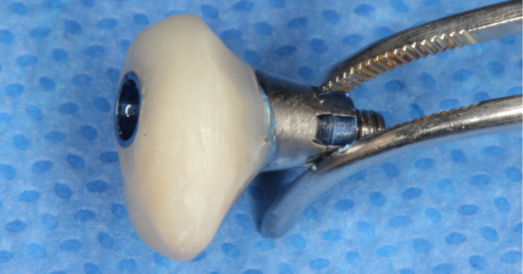
For this reason, my preference is to reserve direct-to-MUA screws for provisional prostheses (as was done with Vortex screws in the example above), while preserving metal-on-metal interfaces for definitive prostheses (i.e., tibases for monolithic zirconia or a titanium bar with a monolithic zirconia sleeve cemented on top).
When efficiency demands responsibility
Two modern realities are shaping restorative implant dentistry. The population is growing and aging, increasing the overall need for restorative care, while the dental laboratory workforce has not expanded at a comparable rate. This imbalance has created a bottleneck in laboratory capacity, contributing to rising laboratory costs. In response, many dentists are taking on a greater share of laboratory-related tasks to manage costs, increasing the pressure to improve efficiency across clinical workflows.
Digital tools such as photogrammetry for dental implants can reduce chair time, simplify appointments, and make more complex care more scalable. However, when advanced workflows become easier to execute, there is a risk that case selection and planning are influenced more by convenience rather than by patient-specific biologic considerations and long-term maintainability.
The responsibility remains with the dentist to select workflows because they best serve the patient’s biology, functional requirements, and long-term restorative needs, not simply because they make the clinical day more efficient.
We must educate patients about alternatives, risks, and benefits; tailor restorative planning to respect and preserve patients’ anatomy whenever possible; communicate clearly with the laboratory about design intent; and take ownership of accurate implant-position capture and the resulting prosthesis fit. These are the types of principles we teach in Spear’s Campus Workshops through hands-on experience, mentorship, and structured training.
If there is one bench-to-chairside lesson worth carrying forward, it’s that accuracy in full arch implant dentistry is not an automatic consequence of digital workflows. It is a clinical responsibility that must be intentionally preserved. Photogrammetry for dental implants can raise the ceiling of what is possible, but only when it is selected thoughtfully, validated rigorously, and used in the service of long-term biologic and restorative success rather than short-term efficiency.
References
- Revilla-León, M., Att, W., Özcan, M., & Rubenstein, J. (2021). Comparison of conventional, photogrammetry, and intraoral scanning accuracy of complete-arch implant impression procedures evaluated with a coordinate measuring machine. The Journal of Prosthetic Dentistry, 125(3), 470-478.
- Ma, B., Yue, X., Sun, Y., Peng, L., & Geng, W. (2021). Accuracy of photogrammetry, intraoral scanning, and conventional impression techniques for complete-arch implant rehabilitation: an in vitro comparative study. BMC Oral Health, 21(1), 636.
- Kosago, P., Ungurawasaporn, C., & Kukiattrakoon, B. (2023). Comparison of the accuracy between conventional and various digital implant impressions for an implant‐supported mandibular complete arch‐fixed prosthesis: An in vitro study. Journal of Prosthodontics, 32(7), 616-624.
- Gómez‐Polo, M., Barmak, A. B., Ortega, R., Rutkunas, V., Kois, J. C., & Revilla‐León, M. (2023). Accuracy, scanning time, and patient satisfaction of stereophotogrammetry systems for acquiring 3D dental implant positions: A systematic review. Journal of Prosthodontics, 32(S2), 208-224.
- Hussein, P. M. O. (2023). Photogrammetry technology in implant dentistry: A systematic review. The Journal of Prosthetic Dentistry, 130(3), 318-326.
- Joensahakij, N., Serichetaphongse, P., & Chengprapakorn, W. (2024). The accuracy of conventional versus digital (intraoral scanner or photogrammetry) impression techniques in full-arch implant-supported prostheses: a systematic review. Evidence-Based Dentistry, 25(4), 216-217.
Frequently Asked Questions
SPEAR campus Workshops
Learn Hands-on From Faculty Who Still Practice
A few days at Spear Campus can change how you diagnose, plan, and present cases, not through more lectures, but through hands-on training led by faculty who treat patients every week. Work through real cases in small groups and leave with techniques you can use on your next patient.
Featured Digest articles
Insights and advice from Spear Faculty and industry experts