Why You Should Do a Diagnostic Wax-Up
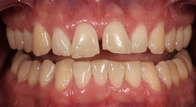
When talking to dentists, I’m frequently asked about when or how to use diagnostic wax-ups (DWUs). This valuable diagnostic tool can be used in your practice to establish a clear visual representation of the desired patient outcome to facilitate treatment planning and communication.
What is a diagnostic wax-up?
It is defined in the Glossary of Prosthodontic Terms as “waxing of intended restorative contours on dental casts for evaluation and planning restorations; a wax replica of a proposed treatment plan. Comparable to trial dentures.”
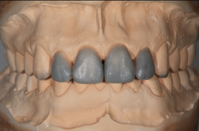
I think of a wax-up as the foundation of the entire treatment plan, because it physically establishes the proposed outcome of esthetics and function. Guides, stents, templates, and matrixes are made on the diagnostic wax-up, or a cast made from the wax-up, to aid in performing surgical and restorative procedures.
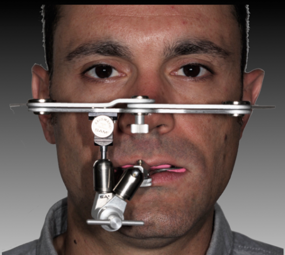
Why you should do a diagnostic wax-up
Because a diagnostic wax-up is an outcome-based diagnostic tool, it should accurately represent the desired result of treatment. The clinician is responsible for accurately communicating on the laboratory prescription the desired outcome the technician is to create with the wax-up.
The instructions written on the prescription form should reflect the needs and expectations of the patient and the clinician. The laboratory’s responsibility is to establish the tooth position, alignment, inclination, morphology, and occlusal scheme based on clinician directions. It should not be arbitrarily done based on the technician’s interpretation of minimal clinical information. The greater the room for interpretation, the more likely clinician and patient expectations will not be met.
Diagnosis and treatment planning with a diagnostic wax-up
By completing a DWU before discussing the proposed treatment plan with the patient, the clinician can confirm that the plan is achievable. It is used as a diagnostic aid to establish the desired esthetic changes (tooth position, alignment, inclination, proportion, and morphology) and develop or confirm the occlusal scheme and function.
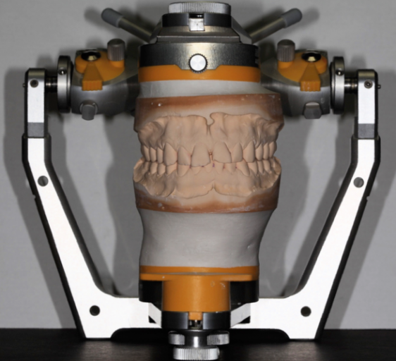
During the waxing process, it may be determined that the plan can’t be achieved from a functional perspective. For example, if you’re restoring the maxillary arch and opening the vertical dimension, the diagnostic wax-up may indicate there is no longer anterior contact with the mandibular anterior teeth. The anterior veneers you had planned or the patient requested won’t work in this situation, because you’d need to add ceramic on the palatal aspect of the upper teeth to reestablish contact with the lower anterior teeth to achieve the desired function.
This necessitates changing the plan from restoring the teeth with veneers to using crown restorations on the maxillary anterior teeth, or doing additional procedures on the mandibular anterior teeth to establish contact.
Pretreatment using a diagnostic wax-up
Some clinicians use the DWU as a visual aid to show the proposed treatment outcome to the patient. Some patients may want to see the proposed esthetic changes in their mouth before accepting treatment. If this is the case, a DWU will provide the basis for creating an intraoral mockup. This step only works well when there are minor changes proposed.
A stent made from the DWU and filled with provisional material (Luxatemp) is placed over the natural teeth and allowed to set. After the stent is removed, the mockup should closely represent the outcome proposed by the DWU. If significant changes in tooth position are proposed, the stent will be distorted when it’s placed over the unprepared teeth that will be changed. Because of this distortion, using a mockup in this situation may defeat its intended purpose.
Using the diagnostic wax-up during treatment
The DWU is used as a template for the desired outcome and is used to create the following treatment tools:
- Two different preparation silicone index/guides. A palatal guide, which includes the incisal edge of the teeth, and a window guide. Their use is critical for ensuring there’s adequate space for the selected restorative material thickness to achieve strength and durability and to create the desired esthetic outcome.
- Pre-preparation: Used to identify tooth surfaces that are out of position when compared with the final restorative outcome. To prevent distortion of the stent, the identified areas must be reduced before creating a mockup.
- During preparation: Used to confirm the depth of the final preparations.
- A copyplast stent. Used to create:
- The intraoral mockup: Pretreatment evaluation and/or mockup for final tooth preparation.
- Provisional restorations: Will be used in trial therapy to evaluate the desired esthetic and functional changes. Any changes made in the provisional restorations should be incorporated into the design and fabrication of the definitive restorations.
- A provisional shell made before the preparation of the teeth.
SPEAR ONLINE
Team Training to Empower Every Role
Spear Online encourages team alignment with role-specific CE video lessons and other resources that enable office managers, assistants and everyone in your practice to understand how they contribute to better patient care.

By: Robert Winter
Date: December 23, 2017
Featured Digest articles
Insights and advice from Spear Faculty and industry experts
