Replacing Adjacent Centrals With Poor Bone Using Implants
By Frank Spear on January 30, 2013 | 0 comments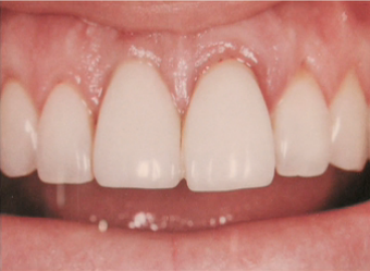
Research has showed us that there are differences in the relationship of the gingival margins and papilla height relative to bone between teeth and implants. Typically the soft tissue is more coronal relative to the bone on teeth than it is on implants.
This is especially true on the interproximal where an average papilla between adjacent teeth may be 4.5 to 5 mm above bone while on adjacent implants the number maybe 3 to 3.5 mm. This makes matching the gingival esthetics of teeth more difficult with adjacent implants. The problem is not as big a challenge on single tooth implants because the bone present on the interproximal of the teeth adjacent to the implant support the papilla at a normal level.
At first glance, this patient who needs to lose both central incisors because of untreatable endodontic problems seems to be pretty straightforward, the papilla heights and gingival margins look reasonably good. However, the patient's radiograph reveals significant bone loss that can make a case like this extremely challenging. Basically the patient's gingiva didn't recede with the bone loss, so she has 7 to 8 mm probing depths around the centrals. What we know before even starting treatment is that because of the bone loss the papilla between the centrals will move apically about 3 to 4 mm from where it is now after we remove the teeth and place the implants.
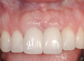
There are really two possible options for solving this problem. One is bone-grafting, removing the teeth, grafting the sockets, and also placing bone to gain vertical height. The procedure has the potential to gain 2 to 3 mm of vertical bone height for the centrals but does not reach that level of success every time. The other option is to consider the use of slow orthodontic extrusion to move the bone more coronally prior to extraction. Unfortunately there are times the bone does not follow the teeth during the extrusion process, especially if there is active infection around the roots as there was in this patient. Ultimately the teeth were extracted, implants placed, and bone and soft tissue grafting performed prior to restoration.
The real key to treating patients like this is controlling their expectations. The likelihood of achieving a perfect result, or even reaching the gingival appearance she had prior to extraction is very low. So it is important to counsel her on the compromises that exist prior to treatment and ideally show her photos of what a predictable outcome with a flat papilla and long contact may look like. In the end this patient was very satisfied with the outcome even though it is less then ideal because of the expectations that were set for her prior to treatment.