Three Key Tooth Positions for Your Orthodontist
By Straty Righellis on June 19, 2012 | 1 comment When upper lateral incisors are missing and the treatment plan is to substitute the upper cuspids to replace the upper lateral incisors, when and how do the restorative dentist and the orthodontist communicate?
If possible, I prefer communication prior to beginning orthodontic treatment to discuss anterior tooth positions. In many cases, the younger patients are still seen by the pedodontist who most likely will not be performing the final restorative work.
In either case, the orthodontist should be aware of three key tooth positions:
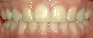
 1. The crown long axis of the substituted cuspid should coincide with the gingival zenith of the substituted lateral incisor. This angulation improvement will allow the restorative dentist to reshape the cuspid to optimal proportions and angulation for maximum esthetics. In addition the orthodontist should position the tooth-gingival margins to provide the “high-low-high” central, lateral, cuspid gingival symmetry as seen in attractive smiles.
2. Intrude the upper bicuspids so the eventual restored bicuspid CEJ will mimic a true cuspid CEJ. This, of course, will position the upper bicuspids out of occlusion until prosthetic buildup is performed.
3. Improve the emergence of the bigger cuspid root to provide improved lingual inclination to the substituted cuspid. Keep in mind this tooth movement requires time as the cuspid has more root surface area. To achieve efficiently, I initially bracket a lower 2nd bicuspid bracket placed upside down on the substituted cuspid to improve the lingual inclination and emergence profile. The lower bicuspid base contour and inclination works well to minimize wire bending.
 About six months prior to the anticipated completion date, the orthodontist and the restorative dentist should collaborate on additional details such as proper anterior spacing for optimal dental proportions for the upper six anterior teeth, and any other details that the restorative dentist may see that the orthodontist may not be aware of.
The combination of photographs and/or models and patient seeing the dentist are beneficial to enhance communication and truly provide collaborative interdisciplinary care.
This communication should include but not be limited to what restorative materials will be used to finalize optimal tooth position and provide the necessary function on the substituted cuspids to provide a mutually protective occlusion. What other tips or ideas do you have? Straty Righellis, DDS. [ www.righellisortho.com ]
Comments
June 21st, 2012